
Bacterial bioerosion of bone may help identify stillborn infants from the past
New research using novel microscopic investigation of bacterial bioerosion of archaeological bone has shown that you can differentiate between stillborn and post-newborn babies. This was most exciting to me as offering a means to contribute to the debate of the interpretation of infanticide in the past, through an investigation of time of death.
Bioerosion is the removal of mineralised substrate through the action of organisms, and has been found to be the most common form of microbial attack of archaeological bone (Figure 1). The author of this new research, Tom Booth from the Natural History Museum, notes that although it was once believed that soil bacteria caused most of this bioerosion in bone, it is the gut microbia that is responsible for corpse putrification that causes this process. Based on the findings that it is the bacteria inside the body that produces this bioerosion, the author thought that this could be useful for assessing different mortuary treatments of the body.
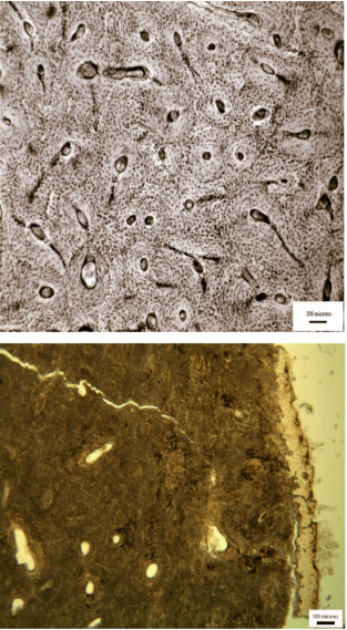
Figure 1: Transmitted light micrograph of a human fresh bone transverse femoral thin section (top) demonstrating perfect microstructural preservation and a typical archaeological femoral section (bottom) where the internal microstructure has been extensively altered by bacteria (from Booth et al., 2015).
To investigate if there is any relationship between bacterial bone bioerosion and funerary treatment, Booth undertook a microscopic analysis of human bones from European prehistoric (4000 B.C. – A.D. 43) and British historical (A.D. 43 – present day) sites. These two assemblages were used as they have been found to have different funerary practices, with the historic period sites practicing burial soon after death, whereas the prehistoric sites have more variable mortuary practices, sometimes including postmortem modification. E.g. Booth and colleagues’ work that found evidence for mummification in Bronze Age Britain using this microscopic method has recently received media attention.
This research shows that irrespective of burial environment, including antiquity or soil type, there was immaculate histological preservation of almost half of the neonatal samples. This is interpreted as the result of sterility of stillborn infant intestinal tracts resulting in the bones being unaffected by the process of bacterial tunneling. In addition, most (12/15) of the unbioeroded newborn samples are from historical cemeteries where most of the other samples had been extensively bioeroded. A previous experimental study by White and Booth using pigs found that bone from stillborn neonatal carcasses had immaculate histological preservation due to the intrinsic sterility of newborn infant intestinal tracts.
Booth found that the soil type had no relationship with bacterial bioerosion. There was evidence for variation in bacterial bioerosion among the later prehistoric assemblages argued to be “consistent with the knowledge that these individuals were subject to variable early post mortem treatment that exposed the bones to diverse levels of bacterial attack.” Bacterial bioerosion in the historical assemblage was high, consistent with that expected within bones of intact bodies that had been interred soon after death.
The use of this novel method to differentiate stillborn vs post-newborn infants can contribute to extending our knowledge of the cause of death during the most crucial time for mother and child in the past, and may also have useful applications for the study of cultural beliefs around stillbirth and post-neonatal death.
References:
Booth, T. J., A. T. Chamberlain and M. P. Pearson (2015). “Mummification in Bronze Age Britain.” Antiquity 89(347): 1155-1173.


