Is this really a 5,000 year old mother and baby?

A recent story of a 4,800-year-old ‘mother’ cradling a baby has been pulling at the heart strings of people worldwide with sensationalist headlines such as “Mother’s enduring love for baby revealed as 5000-year-old fossil found” and “Fossil of 5000-year-old mother cradling baby found in Taiwan”. But is this story everything it’s really cracked up to be?

An archeological team working at a Neolithic site near the city of Taichung since 2014 has unearthed “48 sets of remains”, presumably the number of individual graves, representing the earliest burial site in Taiwan. One of these burials has been described as a mother and baby. However, the news accounts provide little information as to why the researchers believe this to be the case, apart from the placement of the baby with the adult female and the turning of her head to be “looking at her baby” (Figure 1).

Screen Shot 2016-04-29 at 4.44.59 pm
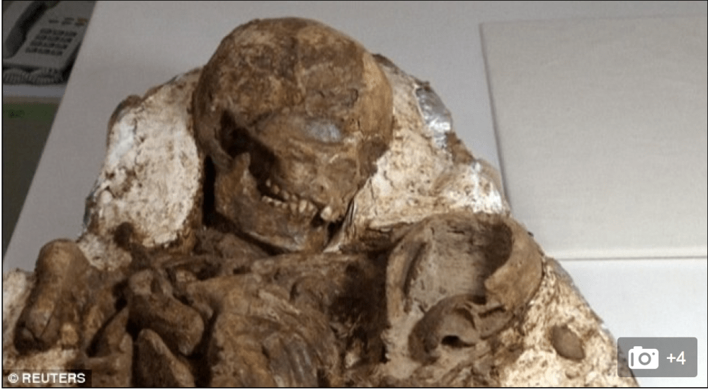
Figure 1: The 4800-year-old “mother and baby” found in Taiwan (source: Reuters)

It is likely that if a female and newborn baby is found in a burial context that they died during childbirth (see my earlier post on fetuses in archaeology). Childbirth is the most critical time for both a mother and baby. This has even led some archaeologists to argue that higher mortality rates of young adult females compared with males represent the hazards of childbirth in the past.

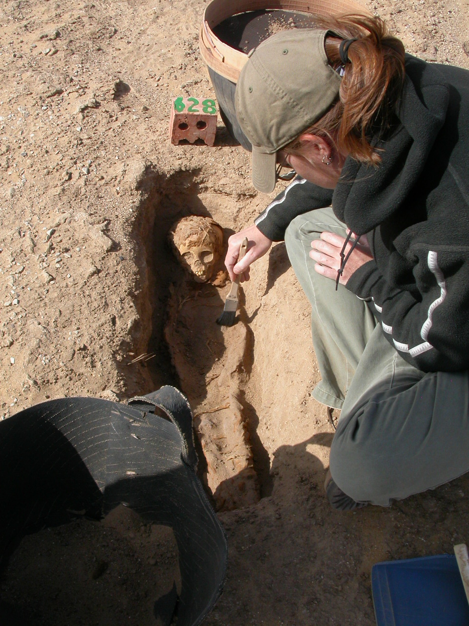
The baby has been described as a foot and a half (about 46 cms), which is about the size of a newborn baby. However, looking at the photos and the videos from the news stories the baby looks too big to be a newborn. The only bones present seem to be from the waist-up. Looking at the relative size of the hands of the archaeologist cleaning the bones and the upper body of the baby (Figure 2), it may be that the size cited is for the upper body, supporting that the infant is older than a newborn. It is difficult to see the cranial bones to assess their development to infer an age-at-death. The cranial bones look thicker than a newborn, but it is unclear as it appears there is some concreted soil adhering to the surface of the bones. Given that this infant seems older than a newborn it is unlikely that they were mother and child.

Screen Shot 2016-04-29 at 2.47.40 pmFigure 2: Archaeologist cleaning the ‘mother-baby’ burial (photo: Reuters video).

In a small Neolithic community there may have been some kind of relationship between the adult female and the infant, or they may have only been buried together because their deaths coincided. Using a cross cultural example, in the Anglican burial tradition babies were interred with non-maternal women in instances of coinciding death (Roberts and Cox 2003: 253).

To assess if there is a biological relationship between this purported mother-baby pair, ancient DNA analyses could be undertaken, but this is difficult with preservation issues in tropical contexts. We should also keep in mind that a mother-child relationship is not always biological.

The fact that the adult female had her head turned to her left may be the result of the burial environment, as some bones can shift in open spaces such as coffins, or from the weight of soil on the bones. Further research looking at the positions of the bone could give more insight on the mode of burial.

We will have to await the scientific presentation of the findings from this site to evaluate the likelihood for this purported mother and baby.

Amazing baby bone facts I: What happens during childbirth

Ever wondered why some newborn babies heads are oblong, resembling ‘cone heads’ after birth and then go back to normal? It is obvious that babies heads are under an enormous amount of pressure during birth, and this is especially so in humans. But how does the actual moulding of the head occur? A newborn baby’s skull bones can move during birth because of the soft tissue between them making the skeletal anatomy somewhat flexible and malleable (figure 1). There are some larger areas of soft tissue between the bones in the front and back portions of the skull called fontanelles.

Screen Shot 2016-04-23 at 8.17.44 am

Figure 1: Childbirth head moulding. From http://keckmedicine.adam.com/content.aspx?productId=117&pid=1&gid=002270

During birth these spaces between the bones allow the baby’s head to change shape. Depending on the amount and length of pressure, the skull bones may even overlap. This overlapping can produce a variety of shapes of the skull from a pointed to a flattened shape (figure 2). This normal moulding generally goes away in a matter of days.

Screen Shot 2016-04-23 at 8.12.54 am

Figure 2: Normal variations of head moulding. From: http://www.open.edu/openlearnworks/mod/oucontent/view.php?id=272&printable=1

These spaces between the bones also allow for the growth of the brain throughout infancy and childhood.

Snap-shots of research: mortuary and biological analyses of fetal, infant and child bodies in Roman Egypt

This month I have the pleasure of showcasing the bioarchaeological work that Dr Sandra Wheeler is doing with fetuses, infants and children in Egypt. Dr. Wheeler is a bioarchaeologist with research expertise in juvenile osteology and mortuary archaeology with a regional focus in ancient Egyptian populations. She is a Lecturer in Anthropology at the University of Central Florida in the United States.

K2 sandra excavating

Dr Wheeler excavating a juvenile burial from the Kellis 2 cemetery, Dakhleh Oasis, Egypt.

Tell me a little bit about your work:

As a bioarchaeologist, I am particularly interested in the synthesis of information gained from the study of the human body as it relates to adaptations and interrelations among the biocultural and natural environments from archaeological contexts. Infants and children are sensitive indicators of environmental and cultural change, so the direct analyses of children’s skeletons and dentitions, as well as the stressors that affected their bodies, provide a unique window into human adaptation to various environments. This, in combination with analyses of mortuary practices, can shed light on cultural ideas of personhood, and child status and agency in past societies. My research aims to interpret patterns of infant and child health and disease to understand the age and risk factors associated with child morbidity and mortality, culture change, and treatment and placement of child bodies at death.

How did you get into your field and why?

I initially began with studies in Mesoamerican archaeology and came to focus on studies of the human skeleton during my Master’s degree. I didn’t know what bioarchaeology was or what it entailed but I knew I wanted to study the human skeleton within its archaeological context. I became interested in the juvenile skeleton and had the wonderful opportunity to illustrate The Osteology of Infants and Children with Brenda Baker, Tosha Dupras, and Matthew Tocheri. This experience strengthened and focused my research interests in juvenile osteology specifically and the archaeology of childhood more broadly. I completed my PhD in Anthropology from the University of Western Ontario in Canada under the direction of Christine White with a focus on bioarchaeology, juvenile osteology and paleopathology, and a regional focus in ancient Egypt. I have been fortunate to work with wonderful colleagues interested in the bioarchaeology of childhood with whom I continue to collaborate and publish. I have had the privilege to conduct fieldwork and publish bioarchaeological research from ancient Egyptian contexts, work that I hope to continue in the future.

K2 analysisDr Wheeler analyzing juvenile skeletal remains excavated from the Kellis 2 cemetery in Egypt.

What is on the future horizon for your research?

I am particularly interested in the full integration of juveniles within bioarchaeological research frameworks, whenever possible. The life course approach is a valuable one for researching trends in stress and disease through time and the risk factors associated with various life stages. I will continue to collaborate with my bioarchaeology colleagues to tease out individual life-histories through the analyses of multiple tissues to better understand the relationships among maternal health, infant survivability, and adult health outcomes, such as the biological and social risk factors for metabolic and infectious diseases.

b.600 3

An example of a juvenile burial from Kellis 2.

Selected publications:

Bleuze, MM, Wheeler, SM, Williams, LJ, Dupras, TL. 2016. Growth of the Pectoral Girdle in a Sample of Juveniles from the Kellis 2 Cemetery, Dakhleh Oasis, Egypt. American Journal of Human Biology. Early View Article first published online: Feb 2016. DOI:10.1002/ajhb.22844. http://onlinelibrary.wiley.com/doi/10.1002/ajhb.22844/abstract

Dupras TL, Wheeler SM, Williams LJ, Sheldrick PG. 2015. Birth in Ancient Egypt: Timing, Trauma, and Triumph? Evidence from the Dakhleh Oasis. In (S Ikram, J Kaiser, R Walker, Eds) Egyptian Bioarchaeology: Human, Animals, and the Environment. Sidestone Academic Press, Leiden pp. 53-65. http://www.sidestone.com/library/egyptian-bioarchaeology

Wheeler SM, Williams L, Beauchesne P, Dupras TL. 2013. Shattered Lives and Broken Childhoods: Evidence of Physical Child Abuse in Ancient Egypt. International Journal of Paleopathology, 3: 71-82. DOI: http://dx.doi.org/10.1016/j.ijpp.2013.03.009

Wheeler SM. 2012. Nutritional and Disease Stress of Juveniles from the Dakhleh Oasis, Egypt. International Journal of Osteoarchaeology, 22(2): 219-234. Article first published online: 2010. DOI: 10.1002/oa.1201.

Wheeler SM, Williams LJ, Dupras TL, Tocheri MW, Molto JE. 2011. Childhood in Roman Egypt: Bioarchaeology of the Kellis 2 Cemetery, Dakhleh Oasis, Egypt. In (M Lally and Alison Moore, Eds.) (Re)Thinking the Little Ancestor: New Perspectives on Infancy and Childhood. Archaeopress, Oxford, pp. 110-121.

Baker BJ, Dupras TL, Tocheri MW. 2005. The Osteology of Infants and Children. Illustrations by SM Wheeler. Texas A&M University Press: College Station.


Short interviews with SSCIP members: Dr Kirsty Squires — sscip

A recent interview that I did with osteoarchaeologist Dr Kirsten Squires for the SSCIP website. Kirsten’s work centres on the treatment of infants and children in the past.

Our interviewee is Dr Kirsty Squires, who is a Lecturer in Osteoarchaeology in the Department of Forensic and Crime Science at Staffordshire University (UK). She has been a member of the Society for the Study of Childhood in the Past since 2011 and is the society’s outreach officer. Tell me a little bit about your … Continue reading Short interviews with SSCIP members: Dr Kirsty Squires

via Short interviews with SSCIP members: Dr Kirsty Squires — sscip