New volume of Childhood in the Past is out now! — sscip

The new volume (9.1) for Childhood in the Past has just been released. Please click the journal link here. Eileen Murphy has provided an editorial that summarises recent SSCIP activities and events, and the great line-up of papers in this volume (below) Editorial Eileen M. Murphy Welcome to the spring issue of Volume nine of […]

via New volume of Childhood in the Past is out now! — sscip

Is this really a 5,000 year old mother and baby?

A recent story of a 4,800-year-old ‘mother’ cradling a baby has been pulling at the heart strings of people worldwide with sensationalist headlines such as “Mother’s enduring love for baby revealed as 5000-year-old fossil found” and “Fossil of 5000-year-old mother cradling baby found in Taiwan”. But is this story everything it’s really cracked up to be?

An archeological team working at a Neolithic site near the city of Taichung since 2014 has unearthed “48 sets of remains”, presumably the number of individual graves, representing the earliest burial site in Taiwan. One of these burials has been described as a mother and baby. However, the news accounts provide little information as to why the researchers believe this to be the case, apart from the placement of the baby with the adult female and the turning of her head to be “looking at her baby” (Figure 1).

Screen Shot 2016-04-29 at 4.44.59 pmFigure 1: The 4800-year-old “mother and baby” found in Taiwan (source: Reuters)

It is likely that if a female and newborn baby is found in a burial context that they died during childbirth (see my earlier post on fetuses in archaeology). Childbirth is the most critical time for both a mother and baby. This has even led some archaeologists to argue that higher mortality rates of young adult females compared with males represent the hazards of childbirth in the past.

The baby has been described as a foot and a half (about 46 cms), which is about the size of a newborn baby. However, looking at the photos and the videos from the news stories the baby looks too big to be a newborn. The only bones present seem to be from the waist-up. Looking at the relative size of the hands of the archaeologist cleaning the bones and the upper body of the baby (Figure 2), it may be that the size cited is for the upper body, supporting that the infant is older than a newborn. It is difficult to see the cranial bones to assess their development to infer an age-at-death. The cranial bones look thicker than a newborn, but it is unclear as it appears there is some concreted soil adhering to the surface of the bones. Given that this infant seems older than a newborn it is unlikely that they were mother and child.

Screen Shot 2016-04-29 at 2.47.40 pmFigure 2: Archaeologist cleaning the ‘mother-baby’ burial (photo: Reuters video).

In a small Neolithic community there may have been some kind of relationship between the adult female and the infant, or they may have only been buried together because their deaths coincided. Using a cross cultural example, in the Anglican burial tradition babies were interred with non-maternal women in instances of coinciding death (Roberts and Cox 2003: 253).

To assess if there is a biological relationship between this purported mother-baby pair, ancient DNA analyses could be undertaken, but this is difficult with preservation issues in tropical contexts. We should also keep in mind that a mother-child relationship is not always biological.

The fact that the adult female had her head turned to her left may be the result of the burial environment, as some bones can shift in open spaces such as coffins, or from the weight of soil on the bones. Further research looking at the positions of the bone could give more insight on the mode of burial.

We will have to await the scientific presentation of the findings from this site to evaluate the likelihood for this purported mother and baby.

Amazing baby bone facts I: What happens during childbirth

Ever wondered why some newborn babies heads are oblong, resembling ‘cone heads’ after birth and then go back to normal? It is obvious that babies heads are under an enormous amount of pressure during birth, and this is especially so in humans. But how does the actual moulding of the head occur? A newborn baby’s skull bones can move during birth because of the soft tissue between them making the skeletal anatomy somewhat flexible and malleable (figure 1). There are some larger areas of soft tissue between the bones in the front and back portions of the skull called fontanelles.

Screen Shot 2016-04-23 at 8.17.44 am

Figure 1: Childbirth head moulding. From http://keckmedicine.adam.com/content.aspx?productId=117&pid=1&gid=002270

During birth these spaces between the bones allow the baby’s head to change shape. Depending on the amount and length of pressure, the skull bones may even overlap. This overlapping can produce a variety of shapes of the skull from a pointed to a flattened shape (figure 2). This normal moulding generally goes away in a matter of days.

Screen Shot 2016-04-23 at 8.12.54 am

Figure 2: Normal variations of head moulding. From: http://www.open.edu/openlearnworks/mod/oucontent/view.php?id=272&printable=1

These spaces between the bones also allow for the growth of the brain throughout infancy and childhood.

Snap-shots of research: mortuary and biological analyses of fetal, infant and child bodies in Roman Egypt

This month I have the pleasure of showcasing the bioarchaeological work that Dr Sandra Wheeler is doing with fetuses, infants and children in Egypt. Dr. Wheeler is a bioarchaeologist with research expertise in juvenile osteology and mortuary archaeology with a regional focus in ancient Egyptian populations. She is a Lecturer in Anthropology at the University of Central Florida in the United States.

K2 sandra excavating

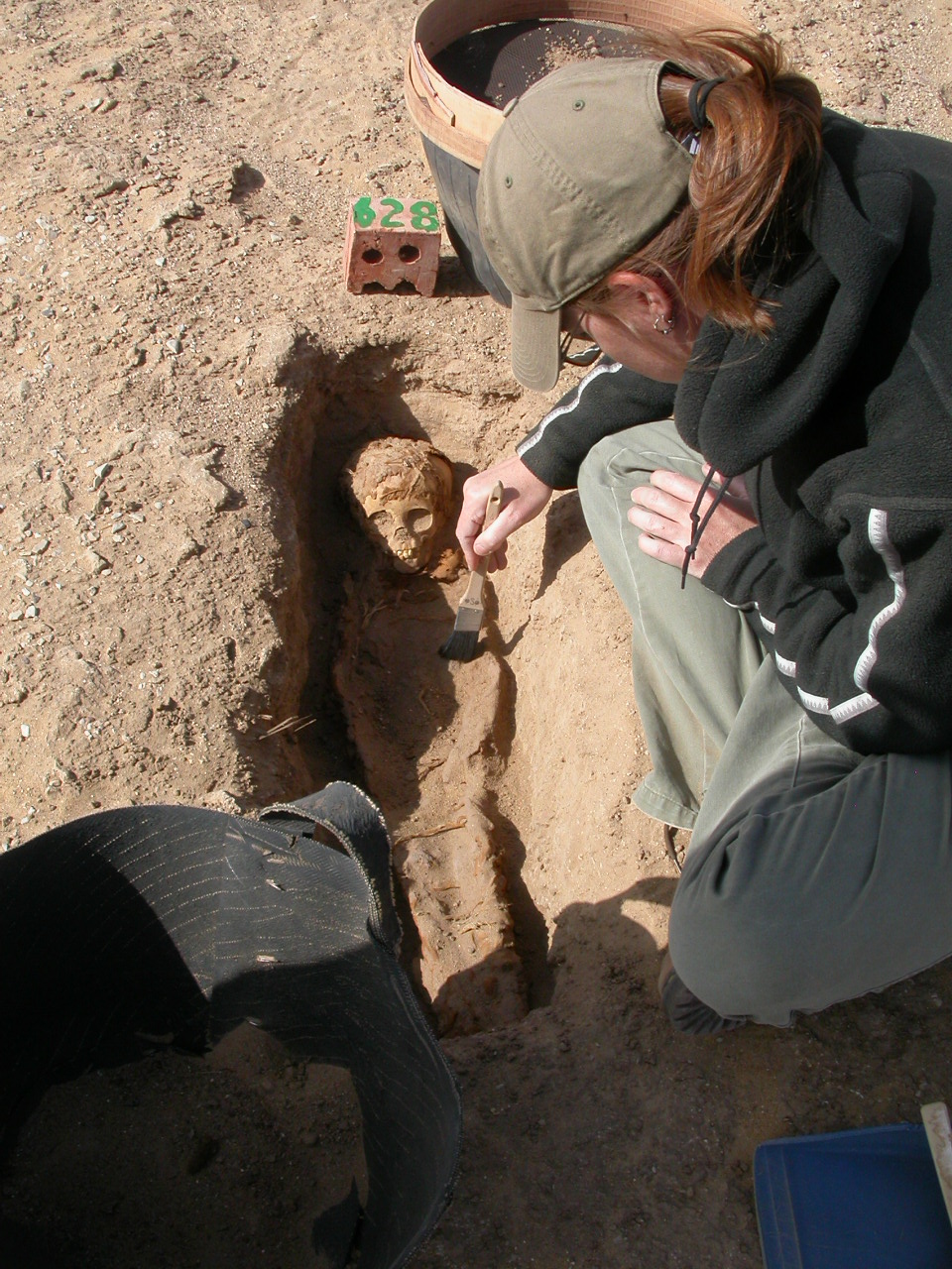
Dr Wheeler excavating a juvenile burial from the Kellis 2 cemetery, Dakhleh Oasis, Egypt.

Tell me a little bit about your work:

As a bioarchaeologist, I am particularly interested in the synthesis of information gained from the study of the human body as it relates to adaptations and interrelations among the biocultural and natural environments from archaeological contexts. Infants and children are sensitive indicators of environmental and cultural change, so the direct analyses of children’s skeletons and dentitions, as well as the stressors that affected their bodies, provide a unique window into human adaptation to various environments. This, in combination with analyses of mortuary practices, can shed light on cultural ideas of personhood, and child status and agency in past societies. My research aims to interpret patterns of infant and child health and disease to understand the age and risk factors associated with child morbidity and mortality, culture change, and treatment and placement of child bodies at death.

How did you get into your field and why?

I initially began with studies in Mesoamerican archaeology and came to focus on studies of the human skeleton during my Master’s degree. I didn’t know what bioarchaeology was or what it entailed but I knew I wanted to study the human skeleton within its archaeological context. I became interested in the juvenile skeleton and had the wonderful opportunity to illustrate The Osteology of Infants and Children with Brenda Baker, Tosha Dupras, and Matthew Tocheri. This experience strengthened and focused my research interests in juvenile osteology specifically and the archaeology of childhood more broadly. I completed my PhD in Anthropology from the University of Western Ontario in Canada under the direction of Christine White with a focus on bioarchaeology, juvenile osteology and paleopathology, and a regional focus in ancient Egypt. I have been fortunate to work with wonderful colleagues interested in the bioarchaeology of childhood with whom I continue to collaborate and publish. I have had the privilege to conduct fieldwork and publish bioarchaeological research from ancient Egyptian contexts, work that I hope to continue in the future.

K2 analysisDr Wheeler analyzing juvenile skeletal remains excavated from the Kellis 2 cemetery in Egypt.

What is on the future horizon for your research?

I am particularly interested in the full integration of juveniles within bioarchaeological research frameworks, whenever possible. The life course approach is a valuable one for researching trends in stress and disease through time and the risk factors associated with various life stages. I will continue to collaborate with my bioarchaeology colleagues to tease out individual life-histories through the analyses of multiple tissues to better understand the relationships among maternal health, infant survivability, and adult health outcomes, such as the biological and social risk factors for metabolic and infectious diseases.

b.600 3

An example of a juvenile burial from Kellis 2.

Selected publications:

Bleuze, MM, Wheeler, SM, Williams, LJ, Dupras, TL. 2016. Growth of the Pectoral Girdle in a Sample of Juveniles from the Kellis 2 Cemetery, Dakhleh Oasis, Egypt. American Journal of Human Biology. Early View Article first published online: Feb 2016. DOI:10.1002/ajhb.22844. http://onlinelibrary.wiley.com/doi/10.1002/ajhb.22844/abstract

Dupras TL, Wheeler SM, Williams LJ, Sheldrick PG. 2015. Birth in Ancient Egypt: Timing, Trauma, and Triumph? Evidence from the Dakhleh Oasis. In (S Ikram, J Kaiser, R Walker, Eds) Egyptian Bioarchaeology: Human, Animals, and the Environment. Sidestone Academic Press, Leiden pp. 53-65. http://www.sidestone.com/library/egyptian-bioarchaeology

Wheeler SM, Williams L, Beauchesne P, Dupras TL. 2013. Shattered Lives and Broken Childhoods: Evidence of Physical Child Abuse in Ancient Egypt. International Journal of Paleopathology, 3: 71-82. DOI: http://dx.doi.org/10.1016/j.ijpp.2013.03.009

Wheeler SM. 2012. Nutritional and Disease Stress of Juveniles from the Dakhleh Oasis, Egypt. International Journal of Osteoarchaeology, 22(2): 219-234. Article first published online: 2010. DOI: 10.1002/oa.1201.

Wheeler SM, Williams LJ, Dupras TL, Tocheri MW, Molto JE. 2011. Childhood in Roman Egypt: Bioarchaeology of the Kellis 2 Cemetery, Dakhleh Oasis, Egypt. In (M Lally and Alison Moore, Eds.) (Re)Thinking the Little Ancestor: New Perspectives on Infancy and Childhood. Archaeopress, Oxford, pp. 110-121.

Baker BJ, Dupras TL, Tocheri MW. 2005. The Osteology of Infants and Children. Illustrations by SM Wheeler. Texas A&M University Press: College Station.
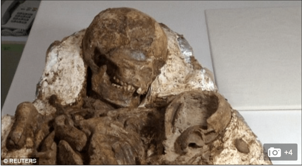


Short interviews with SSCIP members: Dr Kirsty Squires — sscip

A recent interview that I did with osteoarchaeologist Dr Kirsten Squires for the SSCIP website. Kirsten’s work centres on the treatment of infants and children in the past.

Our interviewee is Dr Kirsty Squires, who is a Lecturer in Osteoarchaeology in the Department of Forensic and Crime Science at Staffordshire University (UK). She has been a member of the Society for the Study of Childhood in the Past since 2011 and is the society’s outreach officer. Tell me a little bit about your … Continue reading Short interviews with SSCIP members: Dr Kirsty Squires

via Short interviews with SSCIP members: Dr Kirsty Squires — sscip

Bacterial bioerosion of bone may help identify stillborn infants from the past

New research using novel microscopic investigation of bacterial bioerosion of archaeological bone has shown that you can differentiate between stillborn and post-newborn babies. This was most exciting to me as offering a means to contribute to the debate of the interpretation of infanticide in the past, through an investigation of time of death.

Bioerosion is the removal of mineralised substrate through the action of organisms, and has been found to be the most common form of microbial attack of archaeological bone (Figure 1). The author of this new research, Tom Booth from the Natural History Museum, notes that although it was once believed that soil bacteria caused most of this bioerosion in bone, it is the gut microbia that is responsible for corpse putrification that causes this process. Based on the findings that it is the bacteria inside the body that produces this bioerosion, the author thought that this could be useful for assessing different mortuary treatments of the body.

Screen Shot 2016-03-12 at 9.06.06 pm

Figure 1: Transmitted light micrograph of a human fresh bone transverse femoral thin section (top) demonstrating perfect microstructural preservation and a typical archaeological femoral section (bottom) where the internal microstructure has been extensively altered by bacteria (from Booth et al., 2015).

To investigate if there is any relationship between bacterial bone bioerosion and funerary treatment, Booth undertook a microscopic analysis of human bones from European prehistoric (4000 B.C. – A.D. 43) and British historical (A.D. 43 – present day) sites. These two assemblages were used as they have been found to have different funerary practices, with the historic period sites practicing burial soon after death, whereas the prehistoric sites have more variable mortuary practices, sometimes including postmortem modification. E.g. Booth and colleagues’ work that found evidence for mummification in Bronze Age Britain using this microscopic method has recently received media attention.

This research shows that irrespective of burial environment, including antiquity or soil type, there was immaculate histological preservation of almost half of the neonatal samples. This is interpreted as the result of sterility of stillborn infant intestinal tracts resulting in the bones being unaffected by the process of bacterial tunneling. In addition, most (12/15) of the unbioeroded newborn samples are from historical cemeteries where most of the other samples had been extensively bioeroded. A previous experimental study by White and Booth using pigs found that bone from stillborn neonatal carcasses had immaculate histological preservation due to the intrinsic sterility of newborn infant intestinal tracts.

Booth found that the soil type had no relationship with bacterial bioerosion. There was evidence for variation in bacterial bioerosion among the later prehistoric assemblages argued to be “consistent with the knowledge that these individuals were subject to variable early post mortem treatment that exposed the bones to diverse levels of bacterial attack.” Bacterial bioerosion in the historical assemblage was high, consistent with that expected within bones of intact bodies that had been interred soon after death.

The use of this novel method to differentiate stillborn vs post-newborn infants can contribute to extending our knowledge of the cause of death during the most crucial time for mother and child in the past, and may also have useful applications for the study of cultural beliefs around stillbirth and post-neonatal death.

References:

Booth, T. J., A. T. Chamberlain and M. P. Pearson (2015). “Mummification in Bronze Age Britain.” Antiquity 89(347): 1155-1173.

Violent deaths of children during a time of imperial decline in Ancient Peru

A new study has found that children suffered violent deaths during a turbulent time of severe social stress and drought following the “collapse” of the Wari empire in the Late Intermediate Period (1000-1400 C.E.) in the Peruvian Andes.

A research chapter by Tiffiny Tung from Vanderbilt University and colleagues has shown the locations of fractures predominantly on the left side of the posterior crania strongly suggest that these injuries were not accidental. The severity of fractures with large pieces of bone dislodged indicate that these were formed from violent blows in a standardised manner (Figure 1).

Screen Shot 2016-03-05 at 10.18.08 pm

Figure 1: Top row, Cranium 20: Perimortem trauma on the left side (left photograph) and two perimortem fractures on the posterior (right photograph). Bottom row Cranium 68: perimortem trauma on the left side (bottom left photograph is posterior view, bottom right photograph is posterior-lateral view). Both are from the Late Intermediate Period component at the Vegachayoq Moqo sector at Huari (photo from Taung et al. 2016, figure 10.4, page 207)

The prevalence of these types of trauma in the Late Intermediate Period (n=4/8) is statistically significantly higher than the preceding period where there was no evidence for perimortem trauma in the Wari-era children (N=39).

The authors state that “ … lethal trauma—or any kind of cranial trauma—on children is exceedingly rare in the Andes (except in cases of child sacrifice), so it is particularly revealing of the unstable sociopolitical conditions in the LIP [Late Intermediate Period] in the Ayacucho Basin.”

Previous work by Tung (2008) has shown that more than two thirds of the adults also experienced cranial trauma attesting to a climate of violence during the Late Intermediate Period in this region.

Carbon isotope ratios from enamel apatite and dentine collagen show that maize consumption decreased during this time, and that there was unequal access to maize between the sites, which the authors argue may have led to social tension through inequality.

The authors state that a combination of factors may have contributed to the high level of lethal trauma in the children. The Wari empire represented the first expansive empire in South America, which maintained control of the area from the northern Andes of Peru, to the central Peruvian coast, to the Moquegua Valley in the far south (Schreiber 1992). After its collapse there is evidence for a major transition in social organisation, and increase in competition for scarce resources, supported by the change in patterns of maize consumption. The pattern of trauma found does not suggest child abuse, but rather that the aggressors intended to kill them, argued by the authors to have occurred during raids in the communities.

Although there is no way to definitively tell what caused the increase in lethal trauma in the children, what is clear is that the turbulent times during the imperial collapse likely contributed to their violent deaths.


References:

Schreiber, K. J. (1992). Wari imperialism in Middle Horizon Peru. Ann Arbor: Museum of Anthropology University of Michigan.

Tung, T. A. (2008). Violence after imperial collapse: A study of cranial trauma among Late Intermediate period burials from the former Wari capital, Ayacucho, Peru. Nawpa Pacha, 29, 101–118.


Snap-shots of research: early life stress and later life outcomes in Southeast Asian prehistory

Introducing snap-shots of research in the bioarchaeology of children

As part of this blog, I will feature the work of bioarchaeologists with an interest in infant and child remains through mini-interviews. In particular, I wish to highlight the work of emerging researchers in addition to established researchers. Our first interviewee is Dr Angela Clark, an Early Career Researcher who has an interest child development and morphology.

1) Tell me a little bit about your work?

All living people manage stress. My research addresses important anthropological questions regarding the effects of critical periods in human history, such as the agricultural transition, through examining stress indicators in the bones and teeth. The ultimate size and shape of the adult human skeleton is not only influenced by individual genetic potential, but is a result of the biosocial environment in which a child grew-up. Chronic stress during childhood has significant life-long effects on individual health and population well-being. My research aims to interpret episodes of childhood stress to enhance understanding of human adaptability and variability, and recognise how unique physical environments and sociocultural factors play their role in individual and population health.

2) How did you get into your field and why?

As a teenager I read books by the forensic anthropologist, Kathy Reichs. The human skeleton fascinated me, and I felt particularly drawn to the humanitarian aspect of returning the identity of the living person to the bones, and providing information of the circumstances surrounding an individuals’ death. Since then I have been privileged to examine humans remains, from archaeological contexts from the UK, Thailand, Peru, and the Cook Islands, and in the forensic context in New Zealand.

3) What is on the future horizon for your research?

I am particularly interested in the emerging field of forensic bioarchaeology, integrating my existing research skills with my professional connections and experience in forensic science to extend forensic human identification. My future research will use microscopic methods of human dental enamel to assess individual life-histories of early-life stresses and later health outcomes in both the survivors (adults) and non-survivors (children). From a forensic perspective, these methods can provide a detailed chronology of childhood stress, which can be as evidence in cases of chronic child abuse.

——————————————————————–

Dr Clark is a bioarchaeologist with research expertise in human skeletal and dental developmental plasticity as a response to stress, using a biosocial approach, with a regional focus of Southeast Asia. She is an Affiliate Researcher in the Biological Anthropology Research Group, Department of Anatomy, University of Otago, Dunedin, New Zealand, and is currently coordinating the Forensic Biology Summer School Paper at the University.

Angela’s publications:

Clark, A.L., Tayles, N., Buckley, H.R. and Neuman, F. (2015) The Rima Rau Burial Cave, Atiu, Cook Islands. Journal of Island & Coastal Archaeology, Doi:10.1080/15564894.2015.1050131.
Tayles, N., Halcrow, S. and Clark, A. (2015) Ban Non Wat: Current research on late prehistoric people in the Upper Mun River Valley, Northeast Thailand. In: N.H. Tan (ed.) Advancing Southeast Asian Archaeology 2013. Bangkok: SEAMEO Regional Centre for Archaeology and Fine Arts (SPAFA). pp. 279-288.
Clark, A.L. (2014) Health and sexual dimorphism at Bon Non Wat: the effects of the intensification of agriculture in prehistoric Southeast Asia [Etat de sante et dimorphisme sexuel a Bon Non Wat: Effects de l’intensifiction de l’agriculture dans l’Asie du Sud-Est prehistorique]. BMSAP, 26 (3-4), 196-204.
Clark, A.L., Tayles, N. and Halcrow, S.E. (2014) Aspects of health in prehistoric mainland southeast Asia: Indicators of stress in response to the intensification of rice agriculture. American Journal of Physical Anthropology, 153, 484-495.
Clark, A., Tayles, N. and Halcrow, S. (2012) Sexual dimorphism in adult skeletal remains at Ban Non Wat, Thailand, during the intensification of agriculture in early prehistoric southeast Asia. In: Proceedings of the twelfth annual conference of the British Association for Biological Anthropology


SSCIP Conference Announcement Durham September 2016

The 2016 conference venue and theme for the Society for the Study of Childhood in the Past has just been announced. The theme of Family in Past Perspective will bring together scholars from different disciplines to explore familial relationships through time. Held at the UNESCO World Heritage site in Durham, this conference will not be one to miss!

Source: SSCIP Conference Announcement Durham September 2016