Reflections on the Ethics of Working with Infants from Museum Contexts

I have been reflecting on the work that I have been doing, particularly within museum contexts. There has been a recent increase in interest in the study of the ethics of bioarchaeological practice; however, there has been considerably less in the context of anatomical collections. This is despite the fact that the individuals collected are often from marginalised sectors of the community, e.g., institutionalised individuals.

Within the anatomical museum context, there can be a lack of clear provenance information and loss of relationship between human remains held in collections and acquisition records. However, I feel that to move forward in ethical ways, we need to know who these people were and where they came from. 

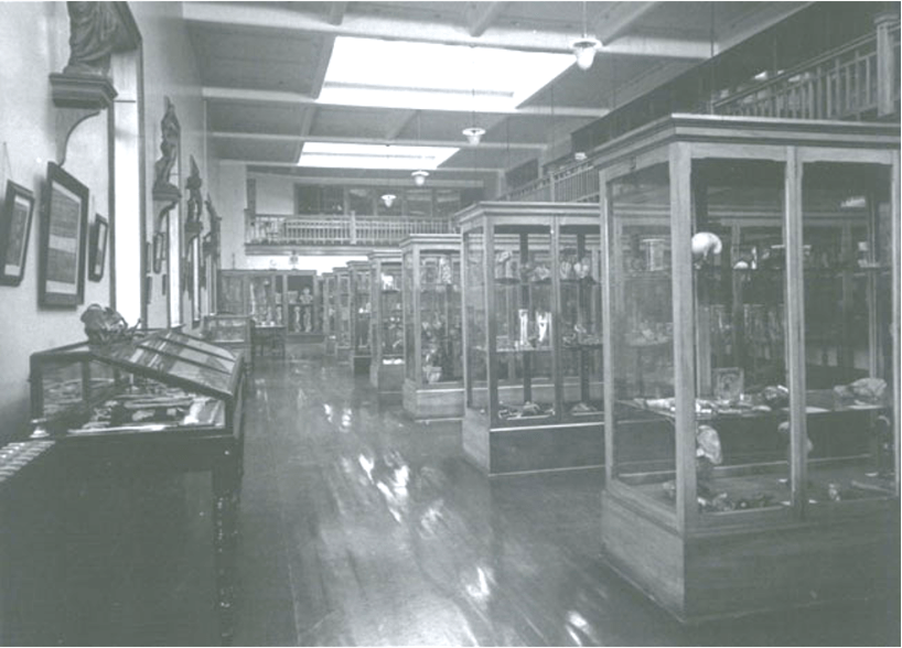
I have, with students and colleagues from Takarangi research, been focused on an analysis of the acquisition records and the skeletal and preserved human remains from the W. D. Trotter Anatomy Museum founded in 1876, which represents the largest anatomical museum in the southern hemisphere. Despite the historical nature of the collection, there is a dearth of research on the people themselves and very little research on the historical archives of acquisition practices.

The Anatomy Museum as it was in 1927
Otago Bulletin

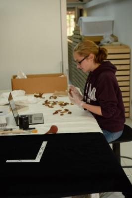
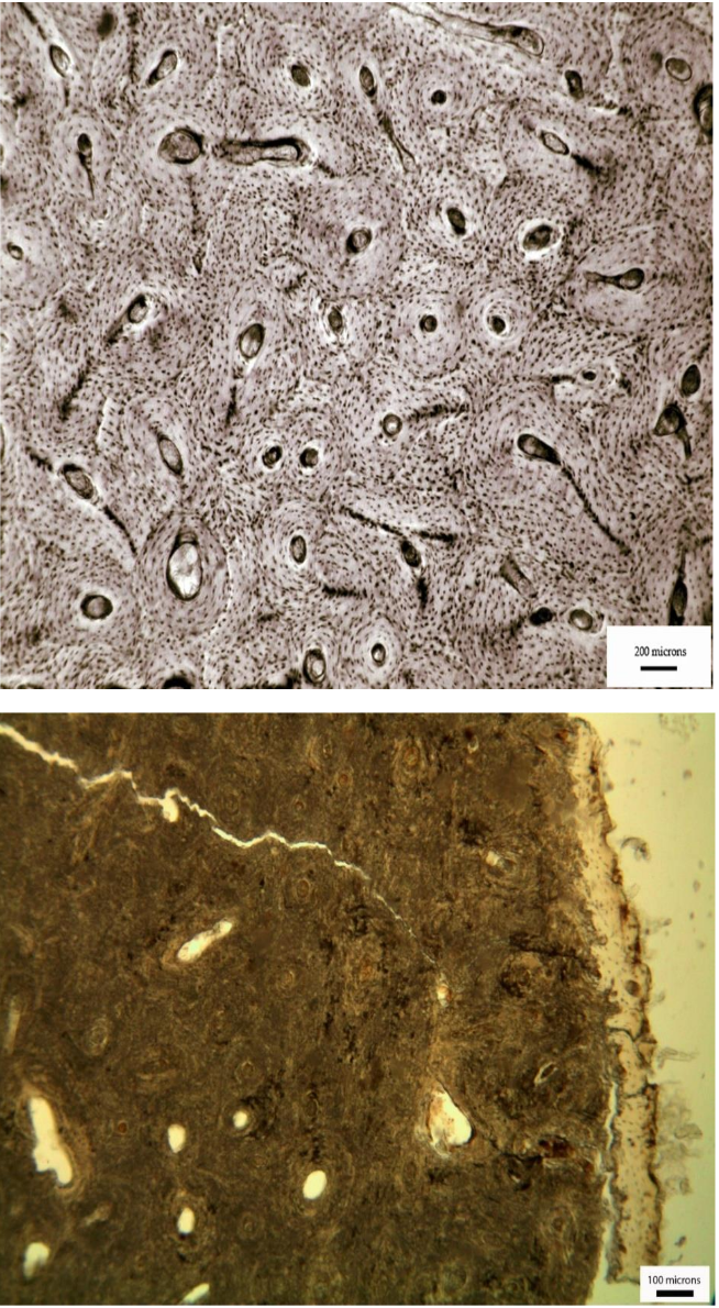
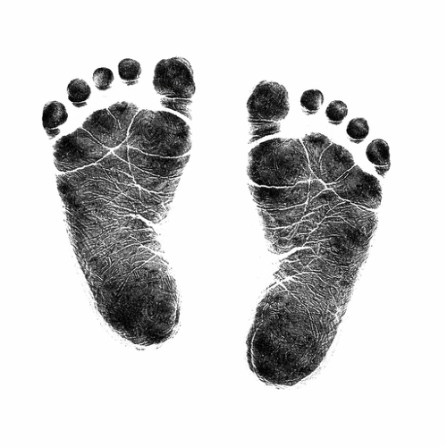
Some of our recent work has looked at the babies represented in the skeletal collections, of which there is a loss of attribution of the acquisition records to the human remains themselves. Through our analysis of the skeletal remains of the infants, we found that there was a number of preterm babies and those with developmental defects. The age-at-death and the pathology reflect the archival records of age and cause of death. The loss of attribution between the records of acquisition and the babies’ remains, along with the way in which a lot of these remains are curated by bone type rather than as individuals, also points to the anatomisation of the body, effectively stripping the individual identity of these babies. We found that of the babies for whom we had names and could find birth and/or death records, many were born to mothers who were unwed and/or from low socio-economic backgrounds. These babies were often born (and died) in mother-baby homes for the unwed (e.g., Batchelor’s Hospital).

The front view of the Forth Street Maternity Hospital (later called Batchelor’s Hospital) in Dunedin. Otago Witness, 18.6.1913

At times, I have questioned whether this research risks repeating the very harm it seeks to address. Some have suggested that analysing these remains may re-objectify the infants, turning them once again into subjects of study. There has also been some hesitation within the university about pursuing this work.

However, I remain of the opinion that silence is not more ethical than engagement. To be transparent about what our collections contain, we must be willing to look closely—however uncomfortable that may be. For me, this work is about re-establishing connection: about naming, contextualising, and acknowledging. It is an attempt, however small, to restore fragments of identity and to confront the histories that allowed these infants to become anonymous specimens in the first place.

This research does not resolve the ethical tensions inherent in working with human remains. But it is, I hope, a step toward greater honesty, accountability, and care.

SSCIP Annual Conference – University of Otago, New Zealand 2021 – Registration and conference schedule

A reminder that the 13th Annual SSCIP Conference is being held via Zoom from the 25th to 28th of October (British Summer Time). It is hosted and organised by Associate Professor Siân Halcrow of the University of Otago, New Zealand, and has been scheduled into eight short sessions over four days to accommodate the different time zones of participants.

Keynote addresses will be made by Professor Maureen Carroll of the University of York, Associate Professor Alison Behie of Australian National University, Professor Holly Dunsworth of the University of Rhode Island, and Professor Sarah Knott of Indiana University Bloomington.

If you are interested in attending any of the eight sessions you can register for free using the following link: https://www.eventbrite.com/e/society-for-the-study-of-childhood-in-the-past-conference-tickets-179748280947. After you register you will be sent a confirmation email. Zoom details for the event can be found by clicking the “View the event” button in this email.

Information regarding the conference schedule and abstracts for all the talks can be found at the following link: https://docs.google.com/document/d/1TqiAhXRo9NEiHPqTD1_OxOgh-wxf4dt0_jcIHQaWG2Y/edit?usp=sharing

Session times:

Day One / 25th October 2021 – Session One: 8am – 10am BST

Day One / 25th October 2021 – Session Two: 9pm – 10pm BST

Day Two / 26th October 2021 – Session One: 8am – 10am BST

Day Two / 26th October 2021 – Session Two: 9pm – 10pm BST

Day Three / 27th October 2021 – Session One:  8am – 10am BST

Day Three / 27th October 2021 – Session Two: 9pm – 11pm BST

Day Four / 28th October 2021, Session One: 8am – 10am BST

Day Four / 28th October 2021 – Session Two: 9pm – 11pm BST

Childhood and the development of urbanisation

The collection of papers in this special issue of Childhood in the Past edited by Francesca Fulminante showcase research on infancy and childhood with sophisticated theoretical and methodological approaches to this topic. This issue represents a significant contribution to understanding the role of children and childhood during the transition to urbanization in Europe through the lens of multiple approaches, including bioarchaeological, archaeological, cognitive developmental (palaeoanthropological), sociological and historical research on infants and children, using a variety of new analytical techniques. This issue moves chronologically from the consideration of cognitive development during prehistory to the nineteenth-century urban environment. Check it out!

Moulded terracotta, beige clay with inclusions, H 9,3 cm, W 6 cm, Veii, sanctuary of Campetti 1, Rome, Museo Nazionale etrusco di Villa Giulia, inv. n. C/168. 3rd–2nd c. BCE. See Pedrucci 2021, 236, Veii 60. Courtesy of the Museum. © Mauro Benedetti.

Why do we have baby teeth?

Baby teeth, also known as milk teeth or deciduous teeth, start forming in the jaws of a baby in utero with the front teeth almost fully formed (apart from the roots) by the time of birth. Baby teeth erupt from about 6 months starting with the front teeth and are usually all present by the age of two and a half years. The first permanent grinding tooth (molar) erupts just behind the last baby molar. Then the front baby teeth get slowly replaced with permanent teeth and by about 12 years of age all the permanent teeth are erupted in the mouth and by adulthood most people have their 3rd molars (“wisdom teeth”).

The jaws of infants and children are far too small to accommodate the larger permanent teeth. Baby teeth are essential for the development of the mouth. They maintain the jaw length, and provide guides for the eruption pathway and therefore proper placement of permanent teeth.

Humans aren’t the only species who have two sets of teeth, but not all animals who have teeth have two sets. Some animals, such as hamsters and moles, only have one set of teeth in their lifetime. Most other vertebrates such as reptiles and fishes have the ability to replace their teeth over and over again. The tooth sizes are very similar and non-specialised.

Screen Shot 2018-02-05 at 7.53.04 PM.png

So, really, the question is why do we only have one set of baby teeth and permanent teeth!

Mammals have very specialised sets of teeth that need to fit together properly to work well. Each tooth has a specific function and they need to work together as a unit, which makes chewing much more efficient for the purposes of getting nutrients from food. If they are constantly being shed and replaced throughout life the precision matching of shape and size of neighbouring teeth that enable that efficient chewing is lost.

But there is a trade off with developing such specialised teeth – it takes more energy to make them, so we are left with one set of teeth during development and one throughout our whole adulthood.


First sighting of a wild newborn chimpanzee taken from mother and cannibalised.

The first ever witnessed case of a newborn wild chimpanzee being taken from the mother and subsequently eaten was published by researchers from Japan this week in the American Journal of Physical Anthropology. The authors explain that this most likely occurred because the pregnant chimpanzee did not go on “maternity leave”, a period near the time of birth that the mother chimpanzee hides and gives birth alone.

Just a matter of seconds after the mother, Devota, gave birth and before she could touch her newborn, an adult male chimpanzee, Darwin, picked up the infant and took it into the bush. The researchers therefore couldn’t confirm if it was a live or still birth or the sex of the infant. Darwin and other members of the group were subsequently seen eating the newborn. At the time of the incident in 2014, there were about 60 members in the group, with 20 adult females and 10 adult males.

Screen Shot 2018-02-03 at 11.55.38 PM.png
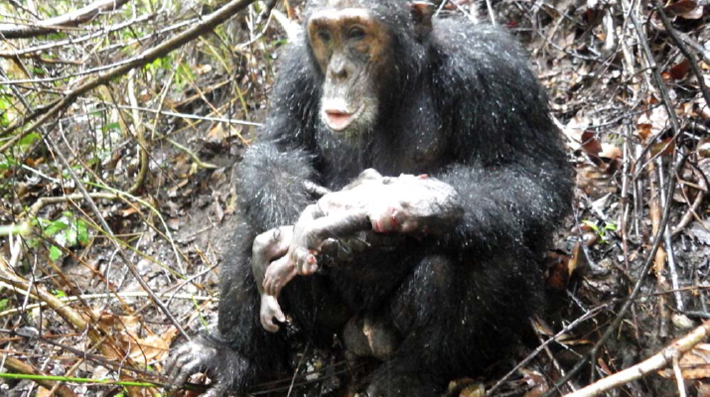
Photo of Darwin with the infant that was subsequently cannibalised (Nishie and Nakamura 2018).

There have been 45 sightings of infanticide in wild chimpanzee groups in six different populations, but never around the time of birth. This incident occurred in group “M” from the Mahale Mountains National Park in Tanzania, which have been studied since 1968. The authors found clear evidence for maternity leave from data from their daily attendance records between 1990 and 2010 with a total of 94 births to 36 females.

Infanticide perpetrated by males has been observed in many primate species and is usually explained as being a male reproductive strategy, whereby males increase their chance of fathering an infant by killing unrelated infants, and results in the mothers becoming fertile again sooner after birth.

The authors propose explanations for why Devota gave birth without “maternity leave”: 1) the baby was preterm and was therefore “not expected”; 2) it was her first pregnancy and had not learned the behaviour of “maternity leave”; and 3) she felt safe in the group as she may have copulated with a number of males around the time of conception of the infant.

Stressed-out mums and demanding children: understanding the maternal – infant interface at the beginnings of agriculture

Modern society is rooted in a dependency on agriculture. Although this is often thought to be a positive human development, the transition to agriculture-based societies had substantial negative impacts on human health, many of which continue to affect millions of people today. The bulk of these negative impacts are borne by the most vulnerable in society – mothers and children.

Recent research in the Arica region in the Atacama Desert in Northern Chile is giving us new insight into how the roots of this transition to agriculture in prehistory affected human society, in particular mothers and their infants. Our research collaboration between the University of Otago in New Zealand, the University of Tarapacá Chile, and Durham University in England is using a multidisciplinary approach to reveal a picture of stresses associated with food shortages, and their possible connections to premature death and vitamin deficiencies in newborn babies.

inca-919101_1920

The Atacama Desert is well-known for the earliest evidence in the world for deliberate mummification of the dead, predating Egyptian mummies by more than two millennia. The intricate funerary rituals associated with the pre-agricultural Chinchorro people of this area were largely focused on infants and children. This has led some to hypothesise that it was a social response to high rates of foetal, infant and maternal death in these populations. Historically, archaeological research in the Atacama has focused on these pre-agricultural mummies, but recent research has highlighted periods of increasing infant mortality later in prehistory – during the transition to agriculture. The ultimate causes of this increase in stress, however, have eluded archaeologists.

Screen Shot 2018-01-28 at 7.17.25 PM.png

The project took a two-pronged approach to this problem, studying changes to diet using chemical signatures in bones and teeth, and assessing their health impacts by looking for signs of pathology on the skeletons of early agricultural populations. Published recently in the International Journal of Paleopathology, and covered here, an Early Formative Period site just transitioning to agriculture (3,600-3,200 years before present) showed that all the infants have evidence of scurvy (nutritional vitamin C deficiency). Interestingly, so did an adult female found buried with her probable unborn child. First author Anne Marie Snoddy says “In addition to contributing to knowledge of the interplay between environment, diet, and health in the Ancient Atacama, this paper provides the first direct evidence of potential maternal-foetal transference of a nutritional deficiency in an archaeological sample”.

This study also used new methods for analysing diet and stress using the chemistry of bones and teeth, these also reveal a picture of early-life stress recently published in the American Journal of Physical Anthropology and covered by Forbes.  “The preservation of mummies in the Atacama gives us an unprecedented opportunity to use tooth chemistry to look at prehistoric infant experience. We have chemical evidence of stress from tissues which form even before the infant is born, showing how the mother’s health is impacting her baby” says author Charlotte King. This work contributes to an understanding of the sensitive relationship between the health of the mother and infant in the past, including the maternal-infant transference of stress signals and micronutrient deficiencies.


Halcrow Anne Marie Snoddy Custom

Anne Marie Snoddy doing her palaeopathological analyses in the Museo Universidad de Tarapacá San Miguel de Azapa, Arica, Chile.


The research is giving new insight into human adaptation to one of the harshest environments in the world. The Atacama Desert experiences less than 2 mm per year of rainfall, making agricultural resources very vulnerable. However, the marine environment is remarkably rich, owing to the upwelling of the cold Humboldt ocean current, resulting in an abundance of marine mammals and fish. Chemical analysis is showing that the people of the desert buffered themselves against the vulnerability of their agricultural resources by continued reliance on these marine foods. Even so, periodic food shortages from El-Niño events in the area were likely, and the skeletal evidence for vitamin C deficiency is interpreted as being related to these events.

A version of this story was originally published here.


Snap-shots of research: Personhood of perinates in the past

This month we are featuring Dr Tracy Betsinger who is an Associate Professor from SUNY Oneonta. Prior to joining SUNY Oneonta, Dr. Betsinger held a post-doctoral research position with the Global History of Health Project at Ohio State University.

1170854_10100934448983597_643943699_n

Tracy working on a perinate from the post-medieval Drawsko collection, Poland (while pregnant with a fetal skeleton shirt on!).

Tell me a little bit about your work:

I’m a bioarchaeologist interested in patterns of health (in general) and infectious disease, particularly treponemal disease, the effects of cultural factors such as status and urbanization on health, and the relationship between mortuary patterning/treatment and identity/personhood, especially among perinates. I work on materials from a variety of contexts, including prehistoric populations from eastern Tennessee and medieval and post-medieval populations from Poland.

How did you get into your field and why?

My interest in perinatal mortuary patterning was a fortuitous happenstance. While working with a colleague, Dr. Amy Scott, on post-medieval Polish materials, we noted the fairly large number of perinatal remains, many of which were well preserved (several with the tympanic rings in place!). We were examining other mortuary patterns at the time, when we decided to investigate the perinatal mortuary pattern to determine whether it matched older subadults or was distinct in some way. We also explored what this might mean in terms of their personhood and identity. The more I began to research perinates, perinatal mortuary patterns, and ontology, the more intrigued I became. I shared my research with a cultural anthropologist in my department (Dr. Sallie Han) whose research is focused on pregnancy and we found much common ground! The result of this was a four-fields anthropology of fetuses, initially an American Anthropological Association session and now a soon-to-be in-press edited volume.

What is on the future horizon for your research?

More recently, I have begun exploring perinatal mortuary treatment with the prehistoric populations from Tennessee. This work is just beginning, but I’m hoping to explore perinatal mortuary patterns/personhood temporally and geographically in the region and dovetail that information about what we know is going on health-wise in East Tennessee. My colleagues (Dr. Michaelyn Harle, Dr. Maria O. Smith) and I have only completed some general assessments of perinates, but so far, there seems to be a consistency in their treatment with older subadults and across time and space. We are planning more nuanced analyses of their mortuary treatment and are hoping to analyze remains for bacterial bioerosion with the hopes of identifying stillbirths from live births.

Bacterial bioerosion of bone may help identify stillborn infants from the past

New research using novel microscopic investigation of bacterial bioerosion of archaeological bone has shown that you can differentiate between stillborn and post-newborn babies. This was most exciting to me as offering a means to contribute to the debate of the interpretation of infanticide in the past, through an investigation of time of death.

Bioerosion is the removal of mineralised substrate through the action of organisms, and has been found to be the most common form of microbial attack of archaeological bone (Figure 1). The author of this new research, Tom Booth from the Natural History Museum, notes that although it was once believed that soil bacteria caused most of this bioerosion in bone, it is the gut microbia that is responsible for corpse putrification that causes this process. Based on the findings that it is the bacteria inside the body that produces this bioerosion, the author thought that this could be useful for assessing different mortuary treatments of the body.

Screen Shot 2016-03-12 at 9.06.06 pm

Figure 1: Transmitted light micrograph of a human fresh bone transverse femoral thin section (top) demonstrating perfect microstructural preservation and a typical archaeological femoral section (bottom) where the internal microstructure has been extensively altered by bacteria (from Booth et al., 2015).

To investigate if there is any relationship between bacterial bone bioerosion and funerary treatment, Booth undertook a microscopic analysis of human bones from European prehistoric (4000 B.C. – A.D. 43) and British historical (A.D. 43 – present day) sites. These two assemblages were used as they have been found to have different funerary practices, with the historic period sites practicing burial soon after death, whereas the prehistoric sites have more variable mortuary practices, sometimes including postmortem modification. E.g. Booth and colleagues’ work that found evidence for mummification in Bronze Age Britain using this microscopic method has recently received media attention.

This research shows that irrespective of burial environment, including antiquity or soil type, there was immaculate histological preservation of almost half of the neonatal samples. This is interpreted as the result of sterility of stillborn infant intestinal tracts resulting in the bones being unaffected by the process of bacterial tunneling. In addition, most (12/15) of the unbioeroded newborn samples are from historical cemeteries where most of the other samples had been extensively bioeroded. A previous experimental study by White and Booth using pigs found that bone from stillborn neonatal carcasses had immaculate histological preservation due to the intrinsic sterility of newborn infant intestinal tracts.

Booth found that the soil type had no relationship with bacterial bioerosion. There was evidence for variation in bacterial bioerosion among the later prehistoric assemblages argued to be “consistent with the knowledge that these individuals were subject to variable early post mortem treatment that exposed the bones to diverse levels of bacterial attack.” Bacterial bioerosion in the historical assemblage was high, consistent with that expected within bones of intact bodies that had been interred soon after death.

The use of this novel method to differentiate stillborn vs post-newborn infants can contribute to extending our knowledge of the cause of death during the most crucial time for mother and child in the past, and may also have useful applications for the study of cultural beliefs around stillbirth and post-neonatal death.

References:

Booth, T. J., A. T. Chamberlain and M. P. Pearson (2015). “Mummification in Bronze Age Britain.” Antiquity 89(347): 1155-1173.