Tiny Bones, Microscopic Insights: A New Look at Neanderthal Babies
How similar were Neanderthals to us, especially at the very beginning of life? Surprisingly, we’ve known very little about how Neanderthal babies grew before birth. That’s mainly because their remains are so rare in the archaeological record.
Now, new research is beginning to fill in that gap.
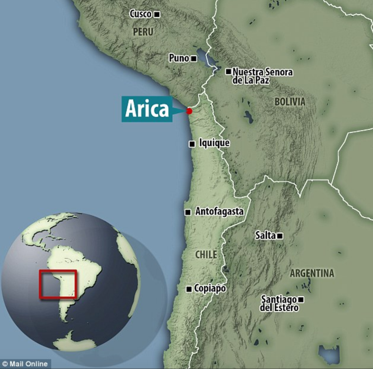
This has taken a closer look literally at the microscopic level of the bones and teeth of three Neanderthal babies. These infants were discovered in the 1960s and 70s at a famous archaeological site called Sesselfelsgrotte in southeastern Germany. The remains date back between 90,000 and 50,000 years.
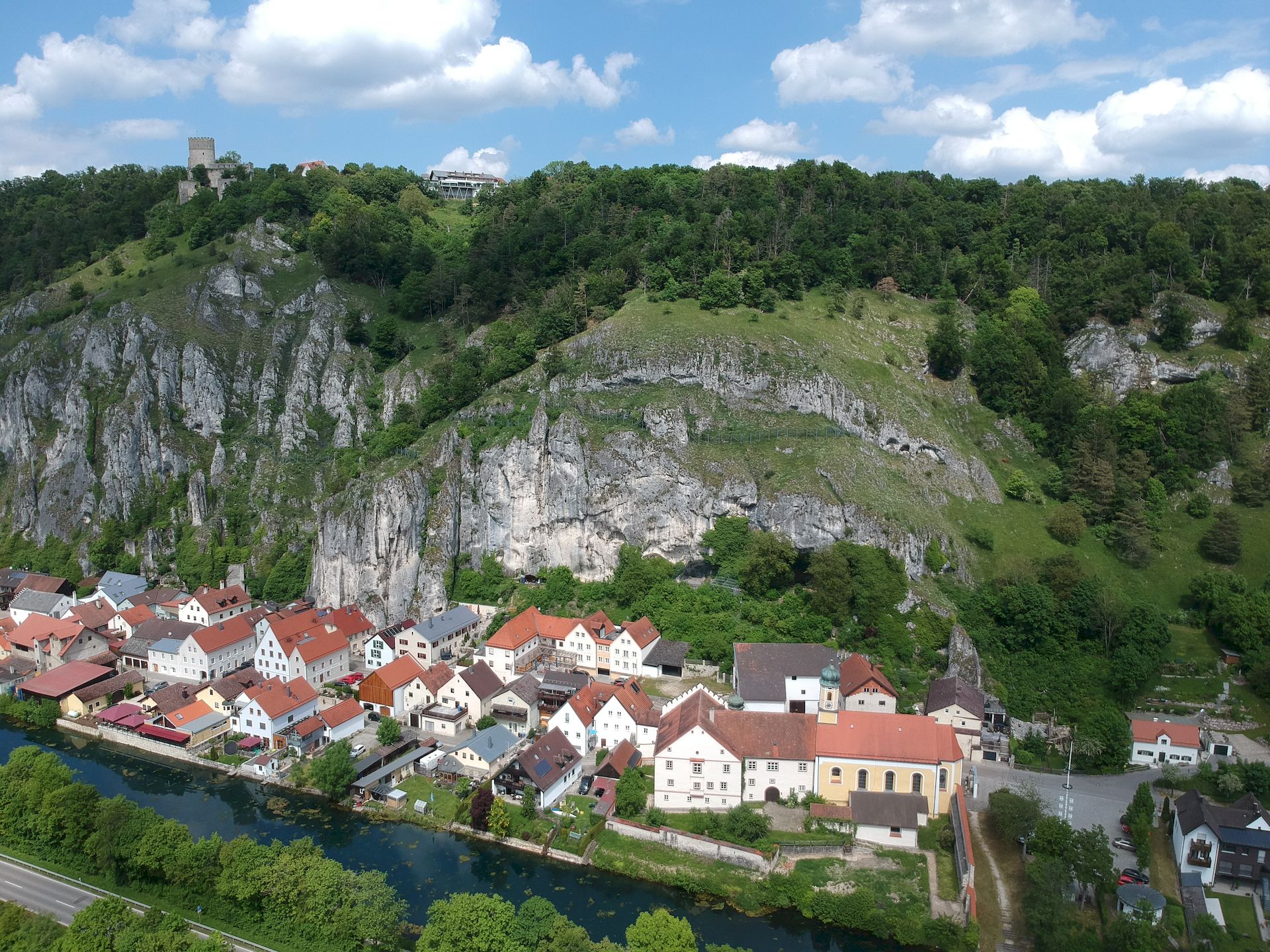
The site of Sesselfelsgrotte, where the cave opening can be seen just behind the large trees above the line of house roofs.
A rare glimpse into Neanderthal infancy
Sesselfelsgrotte is one of the richest Neanderthal sites in western Europe, and what makes it especially remarkable is that it includes the remains of three babies. Finds like this are incredibly rare, making them valuable for understanding early development.
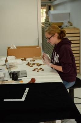
To study the babies without damaging the fragile bones, researchers used advanced micro-CT scanning, a kind of high-resolution 3D imaging. This allowed them to examine both bone structure and teeth in extraordinary detail, and to compare what they saw with how modern human babies develop.
Just like us
What did they find? In many ways, Neanderthal babies developed in broadly similar ways to modern human babies. The microscopic structure of their bones showed clear signs of rapid growth in the later stages of pregnancy.
This adds to growing evidence that Neanderthals and modern humans were closely related, which highlights our shared evolutionary history in a very tangible way.
A surprising clue about ancient health
Inside the teeth, the researchers noticed abnormalities in the dentine—the layer beneath the enamel. These irregularities may point to some kind of metabolic stress during development.
This could represent the earliest evidence of metabolic disease ever found in Neanderthals, dating back around 75,000 years.


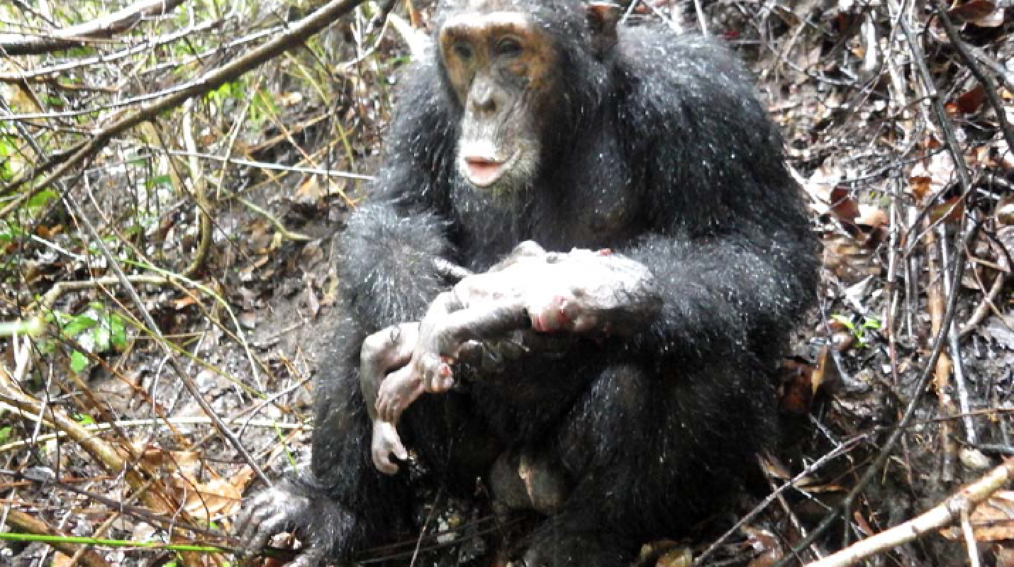 Photo of Darwin with the infant that was subsequently cannibalised (Nishie and Nakamura 2018).