A cursory look through the bioarchaeological literature for explanations of infant death in the past may leave you with a view that infants were being purposefully killed and buried in community cemeteries or simply tossed away in high numbers (e.g. Mays 1993, Mays and Eyers, 2011, Smith and Kahila, 1992).
But what is the likelihood that these accounts are accurate? Here I want to take an analytical look at the bioarchaeological evidence and arguments for infanticide. Some of my views on childhood in the past have been criticised for being clouded by my status as a mother within a ‘Western’ culture. Sometimes I feel that my interpretations are dismissed and put down to my ‘personal’, ‘irrational’, ‘hyper-emotional’, ‘ethnocentric’ thoughts on infancy. One example of this type of experience was at a conference when a senior academic after viewing my poster ‘mansplained’, “you must realise that childhood wasn’t a rosy experience like it is now where you come from. They weren’t wrapped up in cotton wool!”. I wonder if he would have said that if my infant wasn’t attending the conference, the result of no childcare options for participants (dockristy touches on this issue of inclusive conferences for caregivers in her recent blog post).
Infanticide is the intentional killing of infants. Legally, “infanticide” can refer to the deliberate killing of any infant under the age of 12 months (Kellet, 1992). Here I use the term for intentional infant killing around the time of birth, as this is the time in which it usually occurs. Infanticide has been practised in a wide range of cultures through time, and has been argued in some anthropological texts to be an adaptive strategy to environmental, economic, and social circumstances since the Pleistocene era (Hausfater and Hrdy, 1984:xxix).
Common methods for disposing of unwanted children in non-Christian cultures were exposure or drowning without subsequent burial or with covert burial (reviewed in Gilmore and Halcrow, 2014). Some of the motives documented for infanticide include poverty, and if a baby was born with a physical deformity or was “weak”. The sex of infants was also an important factor in infanticide practice for many cultures.
What evidence are these bioarchaeological studies using to inform their interpretations of infanticide? For most papers their main evidence cited for infanticide is a peak rate of mortality around the age of full-term gestation (the perinatal period of about 38-41 weeks gestation) (e.g. Mays 1993, Mays and Eyers, 2011, Smith and Kahila, 1992).*
However, we know from modern age-at-death information that it is normal to see a high rate of infant death at around full-term gestation (see Halcrow et al. 2008 for a review of this evidence). Birth is the most crucial time in a baby’s and mother’s life. Birth and the first few days of life are a dangerous time for a baby with the risk of mortality being extremely high (Kelnar et al., 1995:1). Birth complications, maternal health factors and the risk of disease are likely to have increased the incidence of perinatal deaths and stillbirths in the past. Postpartum dangers include trauma, pneumonia due to infection of the amniotic cavity (Redfern, 2007:185), and respiratory distress syndrome, particularly for pre-term or low birth-weight perinates, owing to the immaturity of the lungs. Environmental hazards for the newborn include infections, bathing in contaminated water, and tetanus due to the use of dirty instruments (Kelnar et al., 1995:6-8, Redfern, 2007:185).
Unsurprisingly, this high rate of infant death around the time of birth has also been found in the archaeological record throughout the world and during different time periods. In the majority of the prehistoric Southeast Asian sites I have worked on we find a high peak of mortality occurring around the time of birth. Other sites with this type of age distribution include Argolid in the Aegean (Angel, 1971), Roman period Britain (Mays, 1993), Southeast Europe (Boric and Stefanovic, 2004), mediaeval and post-mediaeval England (Lewis and Gowland, 2007), post-contact indigenous populations in North America (Owsley and Jantz, 1985), Roman period Egypt (Tocheri et al., 2005), and many more. Were all these cultures at these different time periods killing their infants and then burying them overtly within community cemeteries? I think not. I am not arguing that infanticide never existed in the past. However, these were often discrete events with the dead babies disposed of covertly.
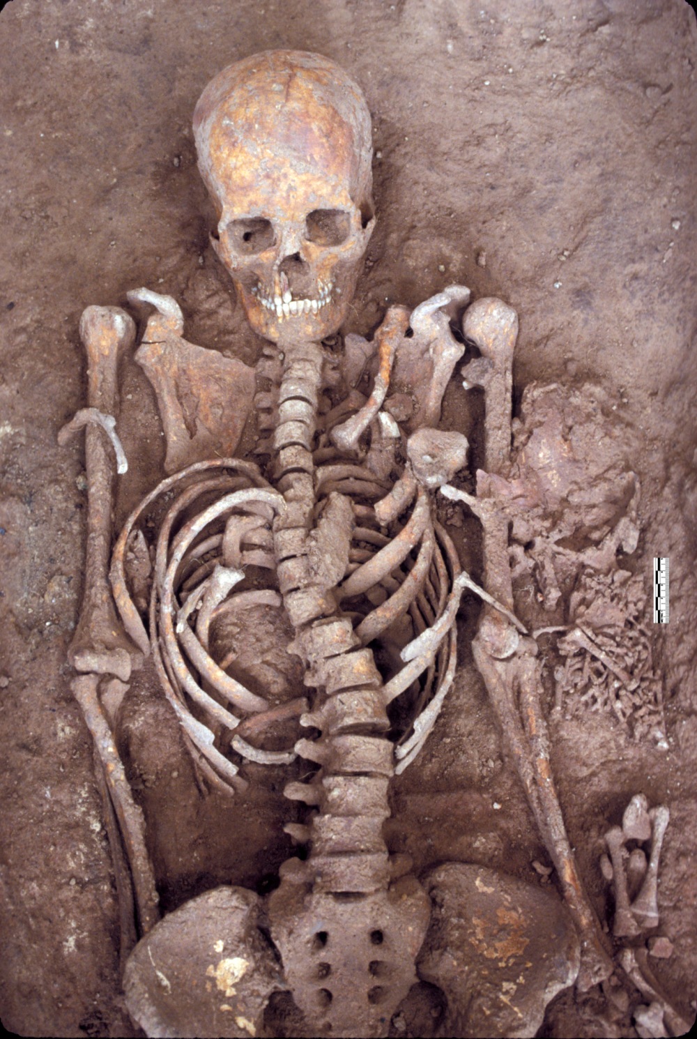 Probable mother and newborn death from the ‘Neolithic’ site of Khok Phanom Di, Southeast Thailand. This site had a infant death representation of over 40% of the cemetery sample.
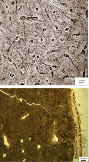
 A ‘foetal’ (preterm) birth from the site of Ban Non Wat, Bronze Age, Northeast Thailand. If a live birth, this baby wouldn’t have lived for long after birth because of its immaturity.
One of these bioarchaeological papers that has interpreted the practice of infanticide is based on the Yewden Roman villa site at Hambleden in Buckinghamshire, England, which became somewhat of an archaeological “celebrity”, showcased by the BBC in 2010 (Mays and Eyers, 2011). The Hambleden site has been identified as a sophisticated “two corridor” Roman villa (Percival, 1990:531). It was first excavated in 1912 by Alfred Heneage Cocks, who reported the discovery of 103 burials, 97 of which were small infants, buried under courtyards or walls on the north side of the site (Cocks, 1921). The infant bones were recently rediscovered in a museum archive after almost a century.
Mays and Eyers (2011) have compared the perinatal age-at-death distribution pattern to other sites that have been interpreted to have an ‘infanticide’ type mortality profile. Other than that there is nothing in the mortuary or archaeological information to suggest that infanticide was probable. The burials at Hambleden are inconsistent with what is known about Roman infanticide practices. As discussed, exposure or drowning were the most usual methods employed, in which case we might expect to find infant bones as haphazard scatters in middens, remote areas of the landscape, or in wells or waterways as has been the case in Scandinavia (Wicker, 1998:215).
An understanding of the historical and ethnographic information on infanticide practices and burial, the historical or other contextual information associated with the site, infant burial practices, and mortality pattern data information is essential for assessing the likelihood for infanticide. It remains that the most parsimonious explanation for cemeteries with a peak of infant death around full-term are the result of a normal age-at-death pattern.
Why then is there a preoccupation or fascination with this idea of infanticide in the past? Were people in the past seen to be of lower moral status and therefore more likely to kill their babies? Could this continued focus on arguments of infanticide stem from an anthropological legacy of the 19th century of exploring ‘dark’, ‘primitive’ cultures, who were seen to lack intelligence and emotion?
Certainly more critical engagement with the literature on infanticide motives, practices, contextual burial information, and medical literature on the causes and timing of normal infant death offers a good approach to review evidence of infant death in the past. Even a mother with a mind ‘clouded’ by breastfeeding hormones and a ‘rosy’ view of childhood can look at the empirical evidence.
————————————————————-
*Smith and Kahila (1992) also include preterm and post-perinatal infants in their “perinate” age category. The preterm infants probably died a natural death, as is likely without modern medical intervention. In historical and modern accounts, infanticide often occurs soon after birth, so the individuals who died in the post-perinatal period were also less likely to be the victims of infanticide.
NOTE: Part of this blog post has been taken from our work in the following papers (all references cited can be found within these publications):
Gilmore, H. and S. E. Halcrow (2014). Interpretations of infanticide in the past. J. Thompson, M.P. Alfonso-Durruty and John Crandell (eds). Tracing Childhood: Bioarchaeological investigations of early lives in antiquity. Florida: University of Florida Press. 123-138.
Halcrow, S. E., N. Tayles and V. Livingstone (2008). “Infant death in prehistoric Southeast Asia” Asian Perspectives. 48 (2): 371-404.
See also Gowland et al. (2014) who offer an excellent re-evaluation of evidence for infanticide in Roman Britain.
Gowland, R. L., A. Chamberlain, & R. C. Redfern (2014). “On the brink of being: re-evaluating infanticide and infant burial in Roman Britain” Journal of Roman Archaeology Supplementary Series 96: 69-88.

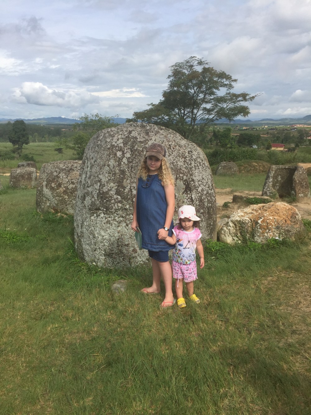 Our visit to the Plain of Jars site 1.
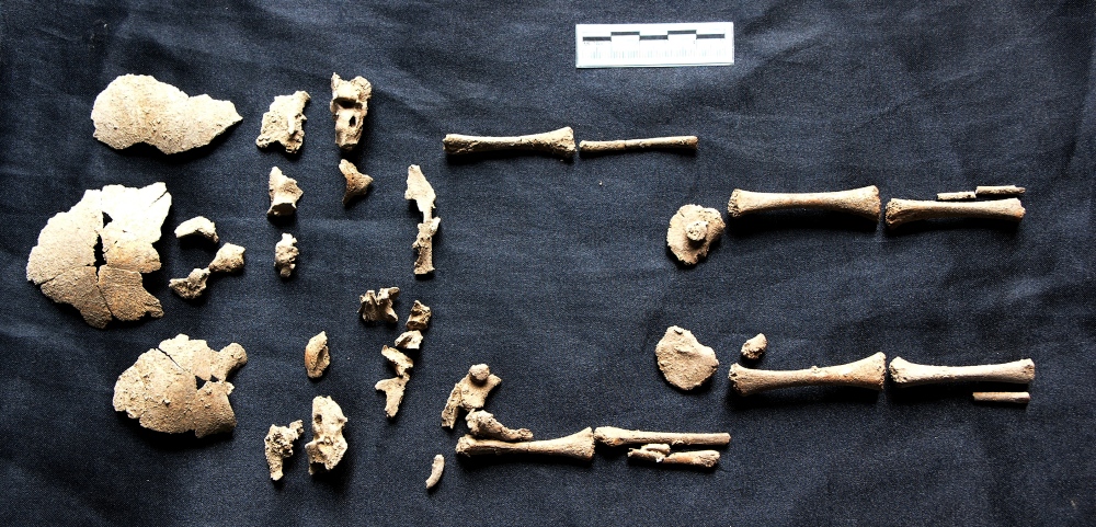
 A 24-26 week old foetus from the Iron Age site of Non Ban Jak, Northeast Thailand.
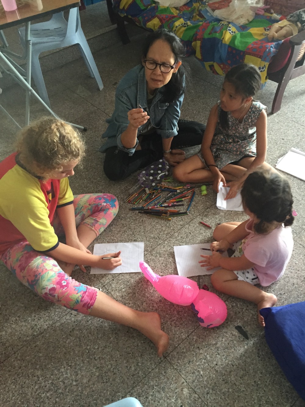 Our “super-nanny”.
 The two-year-old helping me re-box some archeological human remains.
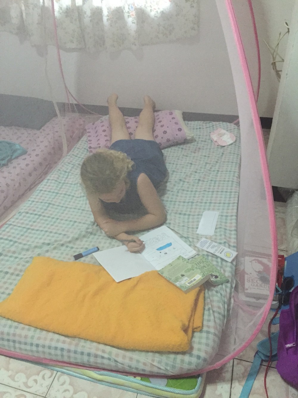 The 11-year-old hiding in our bedroom for some quiet space to do her school work under the mosquito net.


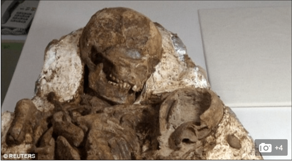 Figure 1: The 4800-year-old “mother and baby” found in Taiwan (source: Reuters)
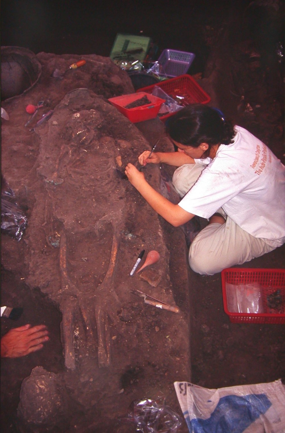
 Figure 2: Archaeologist cleaning the ‘mother-baby’ burial (photo: Reuters video).