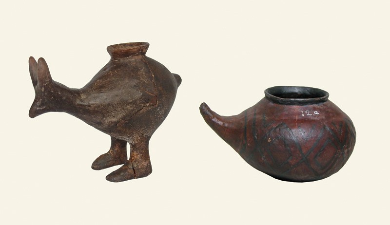
Figure 1 | Ancient pottery vessels. Vessels with a spout for pouring liquid and of a size suitable for feeding babies have been found at archaeological sites. The earliest examples of such vessels5 have been dated to around 5500–4800 bc, but whether these were used to feed infants is unknown. Two vessels are shown of this size and shape from the late Bronze Age or early Iron Age (vessels dated between 1200 and 800 bc). The vessel on the left, from Vösendorf, Austria, is approximately 90 millimetres high. The vessel on the right, from Statzendorf, Austria, is about 85 mm high. Dunne and colleagues’ analysis1 of organic residues found in ancient spouted vessels (not those pictured) sheds light on how early populations might have fed young infants. Credit: Katharina Rebay-Salisbury
For years, many archaeologists ignored children when studying ancient populations, but researchers now increasingly recognize the importance of children when trying to understand the factors affecting earlier societies2,3. One such example concerns a major societal turning point in human prehistory, known as the Neolithic demographic transition, when there is evidence of a substantial increase in fertility and a growth in the number of individuals in human populations compared with that of earlier societies4.
The Neolithic period in Europe began roughly around 7000 bc. During the Neolithic, some humans began to move away from a hunter-gatherer lifestyle towards one that depended on crops and domesticated animals. How did this transition to agriculture lead to a baby boom? An exploration of the approaches used to feed infants might provide some of the evidence needed to answer this question.
Some of the earliest known pottery vessels of a suitable size and shape for use in feeding infants are from the Neolithic period. These artefacts, discovered in Germany, have been dated5 to between 5500 and 4800 bc. It has been suggested6 that during the Neolithic, weaning — when an infant’s diet changes from breast milk to other foods — occurred earlier in an infant’s life than was previously the case. This earlier weaning might have been accomplished by using animal milk and plant sources of carbohydrates. It has been argued that such early weaning could have helped to counteract the period of infertility that can occur while a mother is breastfeeding7, and thus might have led to the increase in fertility and population size during the Neolithic demographic transition. In the archaeological record, this fertility increase is evidenced, somewhat counter-intuitively, by an increase in the number of infants found at burial sites — if more babies are born in a population, then more babies will also die, and be buried8.
Dunne and colleagues examined ceramic vessels with spouts found in children’s graves from burial sites in Bavaria, Germany. One vessel came from a burial site dated to around 1200–800 bc (during the late Bronze Age), and two vessels came from a burial site from around 800–450 bc (during the early Iron Age).
The authors analysed traces of ancient food in these vessels to determine the origin of these residues, by assessing specific characteristics of fatty-acid molecules. Dunne et al. used isotope analysis to study the chemistry of specific compounds in the vessels, and also obtained molecular ‘fingerprints’ of the ancient lipids. They then compared this information with the fingerprints of known reference compounds. This evidence indicates that the vessels contained fatty acids from dairy products, probably milk, that came from domestic ruminant animals. The specific type of animal that provided this milk was not identified.
It is thought that humans first started drinking animal milk in Europe. A study9 published this year of proteins captured in dental plaque provides direct evidence that adults drank animal milk during the Neolithic period in Europe, with the earliest dates for this occurring around 6,000 years ago. Now Dunne et al. present the earliest known evidence of animal milk in small bottles for infants.
The exploration of infant feeding provides information about how babies have been cared for and how social attitudes towards infant feeding have changed over time10. Dunne and colleagues’ investigation of infant feeding during the Neolithic provides insight into cultural beliefs related to the body, infancy and motherhood. Furthermore, the type of food infants are fed, and when during their development they are given food in addition to breast milk, has a strong relationship to infant health and survival11.
Human breast milk is a perfect baby food, containing carbohydrates, protein, fat, vitamins, minerals, digestive enzymes and hormones12. It provides protection from infection because it contains numerous types of immune cell13,14. Some of the sugars it contains, although not digested by babies, support certain communities of gut microorganisms , which prevent disease-causing microbes from establishing a presence in the body14. By contrast, animal-milk products do not provide a complete nutritional source for infants. And the use of hard-to-clean bottles for animal milk poses a risk of exposure to life-threatening infections such as gastroenteritis. The introduction of milk in bottles during the Neolithic, therefore, might have led to a deterioration in the health of some infants.
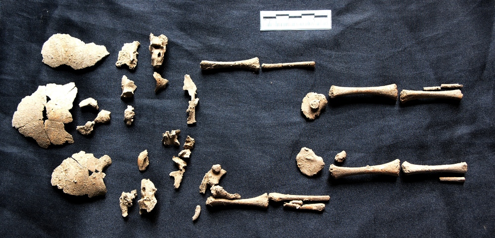
Further research on the remains of people in European prehistoric cemetery sites should be undertaken to explore the effects of the introduction of animal milk as an infant food. This could be assessed by analysing the rate of infant and child mortality, and determining whether any signs of nutritional or infectious disease are present when studying the bones and teeth in infant remains. Furthermore, the age at which a child was weaned can be investigated using techniques that analyse teeth15, and gathering such data can uncover the variation in weaning approaches that existed in a population16. Such knowledge, together with evidence of disease for the individual being studied, might help to provide a greater understanding of the significance of the introduction of animal milk for the lives of ancient children.