Tiny Bones, Microscopic Insights: A New Look at Neanderthal Babies

How similar were Neanderthals to us, especially at the very beginning of life? Surprisingly, we’ve known very little about how Neanderthal babies grew before birth. That’s mainly because their remains are so rare in the archaeological record.

Now, new research is beginning to fill in that gap.

This has taken a closer look literally at the microscopic level of the bones and teeth of three Neanderthal babies. These infants were discovered in the 1960s and 70s at a famous archaeological site called Sesselfelsgrotte in southeastern Germany. The remains date back between 90,000 and 50,000 years.

Essing von der Drohne aus gesehen - in der Bildmitte: Die Felswand mit der Sesselfelsgrotte. Gut sichtbar: Das Stahlgerüst über dem Schutzbau.

The site of Sesselfelsgrotte, where the cave opening can be seen just behind the large trees above the line of house roofs.

A rare glimpse into Neanderthal infancy

Sesselfelsgrotte is one of the richest Neanderthal sites in western Europe, and what makes it especially remarkable is that it includes the remains of three babies. Finds like this are incredibly rare, making them valuable for understanding early development.

To study the babies without damaging the fragile bones, researchers used advanced micro-CT scanning, a kind of high-resolution 3D imaging. This allowed them to examine both bone structure and teeth in extraordinary detail, and to compare what they saw with how modern human babies develop.

Just like us

What did they find? In many ways, Neanderthal babies developed in broadly similar ways to modern human babies. The microscopic structure of their bones showed clear signs of rapid growth in the later stages of pregnancy.

This adds to growing evidence that Neanderthals and modern humans were closely related, which highlights our shared evolutionary history in a very tangible way.

A surprising clue about ancient health

Inside the teeth, the researchers noticed abnormalities in the dentine—the layer beneath the enamel. These irregularities may point to some kind of metabolic stress during development.

This could represent the earliest evidence of metabolic disease ever found in Neanderthals, dating back around 75,000 years.

Check out this upcoming Society for the Study of Childhood in the Past conference

I am looking forward to the range of research to be presented at the 18th Annual Conference for the Society for the Study of Childhood in the Past (SSCIP) on the 10th-12th of June at the University of Oulu, Finland.

This year’s conference theme is on the intersection of childhood, emotions, attachments, and social relationships to uncover how children in past societies experienced and influenced their worlds.

Reflections on the Ethics of Working with Infants from Museum Contexts

I have been reflecting on the work that I have been doing, particularly within museum contexts. There has been a recent increase in interest in the study of the ethics of bioarchaeological practice; however, there has been considerably less in the context of anatomical collections. This is despite the fact that the individuals collected are often from marginalised sectors of the community, e.g., institutionalised individuals.

Within the anatomical museum context, there can be a lack of clear provenance information and loss of relationship between human remains held in collections and acquisition records. However, I feel that to move forward in ethical ways, we need to know who these people were and where they came from. 

I have, with students and colleagues from Takarangi research, been focused on an analysis of the acquisition records and the skeletal and preserved human remains from the W. D. Trotter Anatomy Museum founded in 1876, which represents the largest anatomical museum in the southern hemisphere. Despite the historical nature of the collection, there is a dearth of research on the people themselves and very little research on the historical archives of acquisition practices.

The Anatomy Museum as it was in 1927
Otago Bulletin

Some of our recent work has looked at the babies represented in the skeletal collections, of which there is a loss of attribution of the acquisition records to the human remains themselves. Through our analysis of the skeletal remains of the infants, we found that there was a number of preterm babies and those with developmental defects. The age-at-death and the pathology reflect the archival records of age and cause of death. The loss of attribution between the records of acquisition and the babies’ remains, along with the way in which a lot of these remains are curated by bone type rather than as individuals, also points to the anatomisation of the body, effectively stripping the individual identity of these babies. We found that of the babies for whom we had names and could find birth and/or death records, many were born to mothers who were unwed and/or from low socio-economic backgrounds. These babies were often born (and died) in mother-baby homes for the unwed (e.g., Batchelor’s Hospital).

The front view of the Forth Street Maternity Hospital (later called Batchelor’s Hospital) in Dunedin. Otago Witness, 18.6.1913

At times, I have questioned whether this research risks repeating the very harm it seeks to address. Some have suggested that analysing these remains may re-objectify the infants, turning them once again into subjects of study. There has also been some hesitation within the university about pursuing this work.

However, I remain of the opinion that silence is not more ethical than engagement. To be transparent about what our collections contain, we must be willing to look closely—however uncomfortable that may be. For me, this work is about re-establishing connection: about naming, contextualising, and acknowledging. It is an attempt, however small, to restore fragments of identity and to confront the histories that allowed these infants to become anonymous specimens in the first place.

This research does not resolve the ethical tensions inherent in working with human remains. But it is, I hope, a step toward greater honesty, accountability, and care.

SSCIP Annual Conference – University of Otago, New Zealand 2021 – Registration and conference schedule

A reminder that the 13th Annual SSCIP Conference is being held via Zoom from the 25th to 28th of October (British Summer Time). It is hosted and organised by Associate Professor Siân Halcrow of the University of Otago, New Zealand, and has been scheduled into eight short sessions over four days to accommodate the different time zones of participants.

Keynote addresses will be made by Professor Maureen Carroll of the University of York, Associate Professor Alison Behie of Australian National University, Professor Holly Dunsworth of the University of Rhode Island, and Professor Sarah Knott of Indiana University Bloomington.

If you are interested in attending any of the eight sessions you can register for free using the following link: https://www.eventbrite.com/e/society-for-the-study-of-childhood-in-the-past-conference-tickets-179748280947. After you register you will be sent a confirmation email. Zoom details for the event can be found by clicking the “View the event” button in this email.

Information regarding the conference schedule and abstracts for all the talks can be found at the following link: https://docs.google.com/document/d/1TqiAhXRo9NEiHPqTD1_OxOgh-wxf4dt0_jcIHQaWG2Y/edit?usp=sharing

Session times:

Day One / 25th October 2021 – Session One: 8am – 10am BST

Day One / 25th October 2021 – Session Two: 9pm – 10pm BST

Day Two / 26th October 2021 – Session One: 8am – 10am BST

Day Two / 26th October 2021 – Session Two: 9pm – 10pm BST

Day Three / 27th October 2021 – Session One:  8am – 10am BST

Day Three / 27th October 2021 – Session Two: 9pm – 11pm BST

Day Four / 28th October 2021, Session One: 8am – 10am BST

Day Four / 28th October 2021 – Session Two: 9pm – 11pm BST

Childhood and the development of urbanisation

The collection of papers in this special issue of Childhood in the Past edited by Francesca Fulminante showcase research on infancy and childhood with sophisticated theoretical and methodological approaches to this topic. This issue represents a significant contribution to understanding the role of children and childhood during the transition to urbanization in Europe through the lens of multiple approaches, including bioarchaeological, archaeological, cognitive developmental (palaeoanthropological), sociological and historical research on infants and children, using a variety of new analytical techniques. This issue moves chronologically from the consideration of cognitive development during prehistory to the nineteenth-century urban environment. Check it out!

Moulded terracotta, beige clay with inclusions, H 9,3 cm, W 6 cm, Veii, sanctuary of Campetti 1, Rome, Museo Nazionale etrusco di Villa Giulia, inv. n. C/168. 3rd–2nd c. BCE. See Pedrucci 2021, 236, Veii 60. Courtesy of the Museum. © Mauro Benedetti.

Egyptian ‘hawk’ mummy is a human foetus with a fatal birth defect

Recently researchers have made an unexpected discovery of a mummified foetus while CT scanning a 2300-year-old mummy known as Ta-Kush currently held at the Maidstone Museum in Kent. This coffin was labelled, “A mummified hawk with linen and cartonnage, Ptolemaic period (323 BC – 30 BC).”

Micro-CT scan shows the mummified stillborn human baby. Image: Maidstone Museum UK/Nikon Metrology UK

The high resolution CT scan results have recently been presented at the Extraordinary World Congress on Mummy Studies in the Canary Islands last month. The authors argue that the foetus was about 23-28 weeks gestation and had anencephaly as shown by underdeveloped skull bones.

To me, this begs the question as to whether the several other Egyptian ‘hawk’ mummies curated around the world are actually tiny babies. Further investigation of this baby and others will shed light on the social responses of grief and loss of those born too young to survive.

Watch here on YouTube Mummy ‘bird’ mystery

The coffin. Image: Western University


Snap-shots of research: Personhood of perinates in the past

This month we are featuring Dr Tracy Betsinger who is an Associate Professor from SUNY Oneonta. Prior to joining SUNY Oneonta, Dr. Betsinger held a post-doctoral research position with the Global History of Health Project at Ohio State University.

1170854_10100934448983597_643943699_n

Tracy working on a perinate from the post-medieval Drawsko collection, Poland (while pregnant with a fetal skeleton shirt on!).

Tell me a little bit about your work:

I’m a bioarchaeologist interested in patterns of health (in general) and infectious disease, particularly treponemal disease, the effects of cultural factors such as status and urbanization on health, and the relationship between mortuary patterning/treatment and identity/personhood, especially among perinates. I work on materials from a variety of contexts, including prehistoric populations from eastern Tennessee and medieval and post-medieval populations from Poland.

How did you get into your field and why?

My interest in perinatal mortuary patterning was a fortuitous happenstance. While working with a colleague, Dr. Amy Scott, on post-medieval Polish materials, we noted the fairly large number of perinatal remains, many of which were well preserved (several with the tympanic rings in place!). We were examining other mortuary patterns at the time, when we decided to investigate the perinatal mortuary pattern to determine whether it matched older subadults or was distinct in some way. We also explored what this might mean in terms of their personhood and identity. The more I began to research perinates, perinatal mortuary patterns, and ontology, the more intrigued I became. I shared my research with a cultural anthropologist in my department (Dr. Sallie Han) whose research is focused on pregnancy and we found much common ground! The result of this was a four-fields anthropology of fetuses, initially an American Anthropological Association session and now a soon-to-be in-press edited volume.

What is on the future horizon for your research?

More recently, I have begun exploring perinatal mortuary treatment with the prehistoric populations from Tennessee. This work is just beginning, but I’m hoping to explore perinatal mortuary patterns/personhood temporally and geographically in the region and dovetail that information about what we know is going on health-wise in East Tennessee. My colleagues (Dr. Michaelyn Harle, Dr. Maria O. Smith) and I have only completed some general assessments of perinates, but so far, there seems to be a consistency in their treatment with older subadults and across time and space. We are planning more nuanced analyses of their mortuary treatment and are hoping to analyze remains for bacterial bioerosion with the hopes of identifying stillbirths from live births.

Alien from the Atacama: What baby osteology can tell you

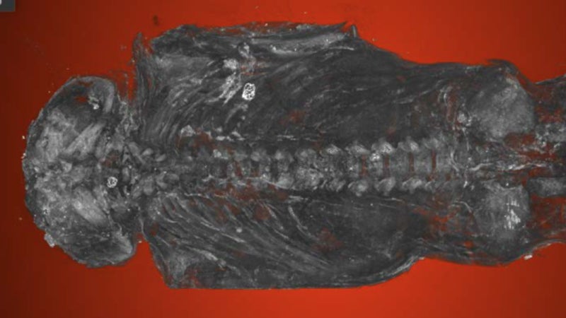
Numerous alien and conspiracy theories have been put forward in the past to explain archaeological finds. One such example that has gained significant media attention is the partially mummified human fetus given the name “Ata” after being found in the Atacama Desert in Northern Chile in 2003. The alien theories and human growth disorder theories that have been put forward are based on the purported unusual skeletal and soft tissue morphology. In 2013, it was reported by geneticist Garry Nolan that the DNA analyses supports that the individual is human. However at this same time it was reported that Ralph Lachman (clinical pediatric radiologist) claimed the skeletal biology was not human-like, citing numerous observations, including “the high level of calcification observed in the legs suggested it was more likely a child between the ages of five and eight years old”.

Screen Shot 2016-06-11 at 9.59.26 pm

Figure 1: Naturally mummified fetus from the Atacama Desert, Northern Chile.

Recently I was approached by a researcher, let’s call him Mr X, who was producing a report from his re-examination of the Atacama specimen. When Mr X asked my opinion to be used in his report he didn’t supply me with any primary data to base my analyses on, so my preliminary observations were based on photos I could find online. Prior to my correspondence with Mr X my colleague based in the UK was asked to comment on the specimen from an ancient DNA perspective. Although the draft report that Mr X emailed for my comments after I had given my preliminary observations concludes that this individual is most likely a human fetus, which I agree with, I was dismayed with a number of things.

Firstly, in this report draft, my colleague’s comments were taken out of context and severely criticized, and included in the report without consent. Perhaps this was because my colleague declined to be sucked into spending precious time and several thousand pounds (things that are not plentiful for scientists these days!) on aDNA analyses of the individual. I should note that my colleague was not worried about Mr X’s criticism of him, but it raised alarm bells for me.

The second issue, and one that I want to discuss here is the lack of proper osteological analyses and reporting, which reminded me somewhat of Dr Kristina Killgrove’s Who Needs an Osteologist installments. Mr X asked me to comment on Mr Z’s (human anatomist and embryologist) interpretations of his findings before writing the report. Mr X advised me to keep the report confidential, as this was being prepared for the private ‘owner’ of the remains based in Spain. The ownership of archaeological remains is problematic in itself. While Mr X had perfectly valid interpretations, a human osteologist’s input is needed for valid scientific analyses of human bone, methodological description and interpretations of the findings. I saw no explanation of age estimation methods, no reference to any human osteological developmental texts, and no inclusion of any studies of mummified soft tissues. As well as bad reporting, Mr X did not acknowledge my input into his findings.

Although I am not going to release the contents of the report, I want to share with you some of my communications with Mr X. Here are some of my explanations of previous biological ‘anomalies’ argued to exist in the Atacama specimen.

1st ‘anomaly’: The 11th and 12th pair of ribs seem to be missing in the radiographs.

My response: The ribs may not be visible in a radiograph as the 11th and 12th ribs are smaller ‘floating’ ribs in that they do not articulate anteriorly at the sternum, are not as robust, and are shorter that the other ribs. There is little information about the formation of ribs in-utero and the timing of the primary centres of ossification (where they first start forming as bone). Initial formation of the 5th-8th ribs start at about 8th-9th weeks in-utero (Scheuer and Black 2000: 238). Scheuer and Black (2000: 238) also state that “by the eleventh and twelfth weeks of intra-uterine life, each rib (often with the exception of the twelfth)”, which implies that the lower ribs are later forming, so may not be as visible in a radiograph.

2nd ‘anomaly’: The seemingly advanced stage of epyphiseal union of the femur, suggesting an age of 5-10 years.

[epiphyseal fusion refers to when the shaft of the bone and the extremity fuse together when the bone stops growing in length]

My response: The statement of the advance stage of epiphyseal fusion is incorrect. If there was fusion/union at the distal femur (which I am assuming they are talking about) this would suggest an adolescent, and thus older than 5-10 years. Regardless of this error in age estimation from epiphyseal fusion methods, I do not see evidence for union on the radiograph online – where is the ‘density’ that they are referring to? There is no ossification of the epiphyses (the unfused extremities of the femora or tibiae) to suggest that fusion of the diaphyses (shaft) and the epiphyses (extremity) would be possible. These bones and the development of these bones all look normal from my observations of the photos and radiographs online.

3rd ‘anomaly’: The epiphyseal plate x-ray density test for age determination suggested an age of 6-8 years old.

My response: This type of age estimation is problematic, and I don’t know any bioarchaeologist or forensic anthropologist who uses the method described. This can’t be applied to mummified remains if it relies on water density.

This is no alien. This was the result of a mother losing her baby early during her pregnancy in the past in South America.

474610_10151344355967437_48598741_o

Figure 2: My archaeologist colleague on our trip to an archaeological site in Arica region, Atacama desert, Chile.

Also see my post on human fetuses in the past here.