Motherhood and Feminism are not Dirty Words: Reimagining Archaeological Practice
For International Women’s Day, I wanted to reflect on some of my old and new writings on the intersection of motherhood and archaeology.
Gendered and intersectional inequities shape access, safety, and participation within field‑based research in archaeology and biological anthropology. Compounding these issues, mothers* routinely confront discrimination and structural barriers associated with pregnancy, breastfeeding, childcare, and norms embedded in fieldwork culture. I have written on some of these issues here on challenges of research and fieldwork with children, attending conferences, and sexism in academic archaeology.
Systemic barriers place disproportionate pressure on mothers compared with fathers, contributing to reduced working hours and widening gender pay gaps (Kleven et al. 2018). The “leaky pipeline” in STEM—arguably a burst main—is strongly linked to caregiving responsibilities; evidence suggests more male STEM leaders have children compared with female leaders (McCabe et al. 2024). Although biological anthropology (including bioarchaeology) is numerically dominated by women, inequities persist in leadership, conference participation, and grant funding (Casad et al., 2022; Turner et al. 2018). Fieldwork remains a particularly acute site of inequality, with persistent reports of discrimination, harassment, and complex logistics for pregnancy, breastfeeding, and childcare (Camp, 2019; Hodgkins & Thompson 2022).
Archaeology’s professional identity is closely tied to fieldwork. Extended time away from home, the physical demands of excavation, and colonial “frontier” narratives have long shaped a disciplinary culture associated with masculine ideals (Tomášková 2007; Moser 2007). In 2013, the Society for American Archaeology established a Task Force on Gender Disparities in Archaeological Grant Submissions; the 2017 report found that nearly all women cited conflicts between fieldwork and parenting that negatively affected their grant submissions (Goldstein et al. 2018). While geography has more thoroughly addressed parenthood and fieldwork (Hope et al. 2019; Jenkins 2020; Price & Hall 2024), archaeology is only recently building momentum through personal accounts, op‑eds, and blog posts (e.g., Halcrow 2017; Hodgkins & Palmer; Norton 2025; Hoag & Von Jena 2025). This expanding body of narrative scholarship coincides with a stagnation in formal gender‑equity research in archaeology (Tomášková 2007; Fladd et al. 2026; Moen 2017; Wylie 2007).
Centring Mothers in Feminism
O’Reilly (2021) argues that despite the diversification of feminist theory—such as queer feminism, third‑wave feminism, womanism, ecofeminism—academic feminism has not sufficiently centred the specific needs of mothers. This omission has contributed to the conflation of mothering and motherhood; to misreadings that equate matricentric feminism with gender essentialism or maternalism; and to the rise of postmaternal thinking and “radical forgetting,” whereby earlier maternal‑oriented activism is dismissed in favour of a degendered feminism (hooks 1984; Stephens 2016; O’Reilly 2021). Based on decades of research and conversations with mothers, O’Reilly (2021) contends that mothering is central to many women’s identities and must be integrated into gendered models of society.
Accordingly, matricentric feminism “puts motherhood at its centre,” treating mothers, mothering, and motherhood as topics deserving sustained inquiry and as a basis for research and activism that contest oppressive institutions and envision empowering maternal identities and practices (O’Reilly 2021).
I have started to use personal narrative as disciplinary critique within a matricentric feminist lens. Caring responsibilities generate distinct vulnerabilities but also unique insights and forms of relational engagement in the field. Here are some reflections and recommendations to reconfigure archaeological practice so that it meaningfully includes and empowers mothers and in particular single parents:
Over 20 years, I have encountered the good, the bad, and the ugly. I have faced blatant discrimination—including being removed from a field project due to pregnancy—and I have missed opportunities because of assumptions about motherhood. Yet I have also experienced meaningful support and inclusion. The following reflections synthesise lessons learned:
- Find a mentor who is inclusive, supportive, and able to communicate openly about your needs.
- Motherhood can enrich scholarship. Parenting has deepened my perspectives on embodiment, ethics, and the social worlds of my research.
- Children expand field relations. My children often helped build rapport with communities, facilitating trust and dialogue.
- Collaborate with other parents. See Lozano & Sánchez (2023) for an example of practical strategies for conducting fieldwork as scientist mothers.
- Advocate for equity in fieldwork opportunities within institutions and professional bodies.
- Share your story when possible; narrative accounts help normalise motherhood in the field and push disciplinary boundaries.
- Expand the evidence base. More systematic qualitative and quantitative studies are needed to understand and address inequities.
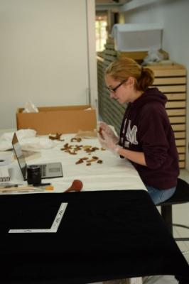
The COVID period, combined with reflections on well‑being and caregiving—including supporting an adult child with disabilities—prompted me to pivot toward collections‑based research, working with human remains already within my department. This shift has produced new grants and collaborations. This trajectory aligns with my ethical commitments and reframes success around work that makes a difference—challenging assumptions about what counts as “fieldwork” and broadening bioarchaeology’s remit to include historically contextualised human remains in collections.
From Personal Narrative to Change: What Societies and Institutions Can Do
Archaeology presents distinctive barriers for parents, and in particular, single parents, because career progression is closely tied to excavation seasons, mobility, and extended time away from home. Academic societies are uniquely positioned to lead practical and cultural change. The following actions outline feasible reforms:
1) Reform field school and excavation expectations
- Accredit alternative training pathways (lab, museum, digital archaeology, community archaeology).
- Recognise local/short‑duration excavations as equivalent experience.
- Encourage hybrid models (e.g., remote recording).
2) Fund childcare and caregiver travel for field seasons
- Introduce seasonal childcare bursaries, not just conference childcare.
- Create grants for caregiver travel and family‑suitable accommodation. Even small funds can make participation viable, especially where field pay is low or unpaid.
3) Publish family-inclusive excavation guidelines
- Standards for family‑safe accommodation, sanitation, and security.
- Predictable scheduling where possible.
- Risk assessments that include dependents.
- Guidance for breastfeeding, pumping, and infant care in field settings. Comparable inclusion toolkits exist in other field sciences; archaeology needs discipline‑wide standards.
4) Build mentorship networks for archaeologist parents
- Cross‑career mentorship programmes.
- Panels with excavation directors who are caregivers.
- Practical guides (e.g., “How I ran a dig with kids”).
5) Support local and community archaeology pathways
- Fund micro‑grants for local projects and distributed collaborations that reduce mobility burdens.
6) Advocate for systemic funding changes
- Lobby for dependent‑care costs to be eligible grant expenses.
- Parental‑leave extensions aligned with excavation seasons.
- Paid field school placements to reduce inequity.
*Following O’Reilly (2021), the term ‘mothers” refers here to those who are doing the mothering and ‘motherwork’, as defined by Sara Ruddick (1989) as maternal practice, and can be undertaken by people other than biological mothers.
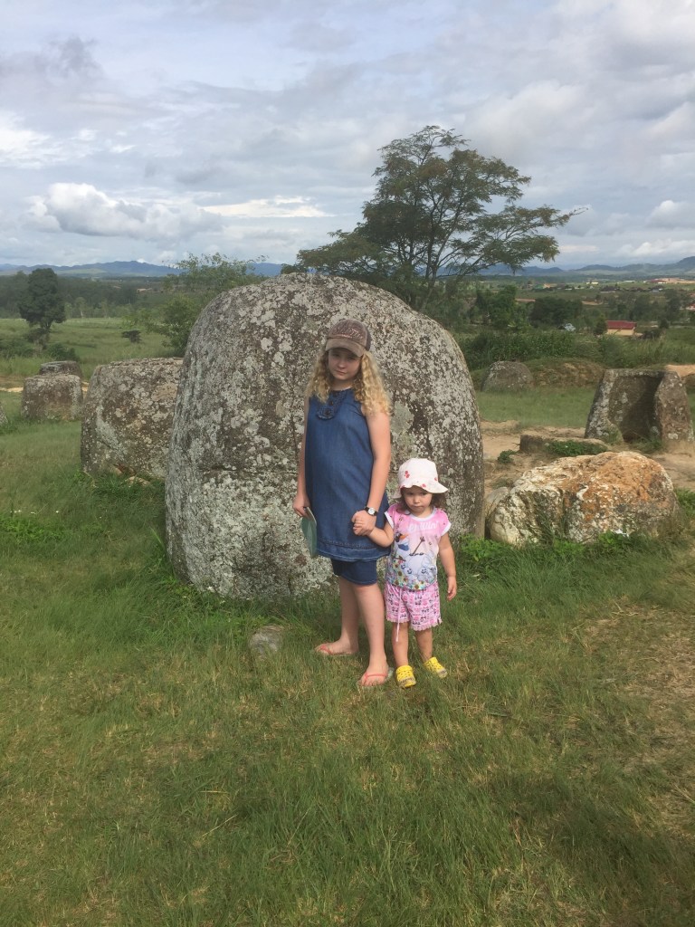
Visit to the Plain of Jars, Laos PDR.
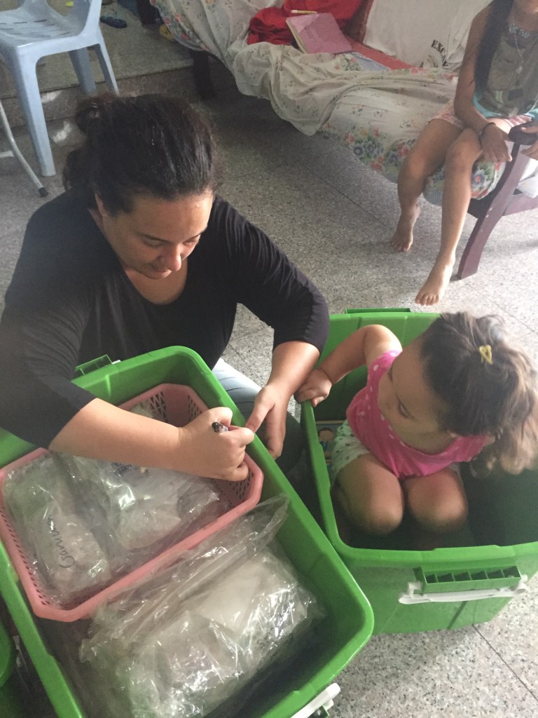
Packing human burials away with some ‘help’