Why were so many babies murdered in the past?

Hundreds of babies of prostitutes getting thrown down a water well in ancient Roman times in Israel; whole cemeteries of unwanted ‘brothel babies’ in Roman period Britain; thousands of Carthaginian babies sacrificed; and purported sacrificial Mayan child victims with ‘supernatural’ obsidian stones. These are just some of the kinds of sensational research stories on infant burials from archaeological collections that are frequently reported. The preoccupation of archaeological research with the subject of infant murder and sacrifice may conjure up images of babies being uncared for in the past, and that infanticide was a common or even accepted practice. However, as with any research, it is important to ask how we can check the validity of these interpretations. Using multi-faceted anthropological studies, we can get closer to disentangling the truth on infant murder in the past.

In legal terms “infanticide” refers to the deliberate act of killing any infant under the age of 12 months. The act of killing unwanted babies is often carried out at the time of birth (the neonatal period), so the term “infanticide” is often used as a synonym for “neonaticide”. It has been stated that babies have been killed in many cultures and in all times in history. Anthropologist Laila Williamson (1978: 61) has gone as far to argue that:

“Infanticide has been practiced on every continent and by people on every level of cultural complexity, from hunter gatherers to high civilizations, including our own ancestors. Rather than being an exception, then, it has been the rule.”


Screen Shot 2017-09-21 at 11.03.57 AM

Figure 1. Anti-infanticide tract depiction of infanticide by drowning, Qing Dynasty, circa 1800.

Non-human primates, including our nearest living relatives, the chimpanzee, have been observed carrying out infanticide. In chimpanzees, this is typically undertaken by an unrelated adult male, the reasoning often hypothesised to be a type of sexual selection to confer reproductive advantage to the male. More recently female-led infanticide has been observed in chimpanzees, the perpetrators also being unrelated to the infants.

The motives for human infanticide are varied. Unique to humans is gender-based infanticide, and it is a parent who often carries out the infant killing. A striking example of gender-based practice is modern female feticide and infanticide, with around half a million female fetuses purposely aborted in India each year alone, as well as the thousands of female babies that are killed soon after birth. Other causative factors for human infanticide relate to poverty, social pressure, and the birth of infants with severe physical deformities. The interplay of poverty and domestic violence towards mothers are argued to have played an integral role in the famous ethnographic research by Scheper-Hughes in which she argued selective neglect or “passive infanticide” occurred in shantytown Brazil.

The actual acts of infanticide in humans are usually non-violent or ‘passive’, including exposure and smothering. The most common method for killing babies in non-Christian societies was drowning. For example, historical texts from the Qing Dynasty often use the term ni nü (to drown girls). There is also documentary evidence for drowning in the Roman Empire, classical Greece, and in Viking Scandinavia. The practice of infanticide is also often carried out covertly and without normative burial ritual.

Although there is documentary evidence for the practice of infanticide in many places and times in the world, most cultures actually condemn its practice, and some would argue that instances of infanticide are generally isolated.

Why, then, is there such a research focus on the practice of infanticide in our past? Do these simply appeal to researchers for publishing a high impact publication, or to news agencies publishing sensational click-bait stories that tug at our heartstrings?

Screen Shot 2017-09-21 at 11.16.47 AM

Figure 2. Early 19th century engraving by an artist associated with William Carey, purporting to show infanticide by drowning in the Ganges River.

Although anthropologists are generally very careful to recognise their own cultural biases in their research, there is undoubtedly a hangover from the 19th century interest of “others” and “dark” practices. Or do anthropologists in recognising their subjective biases on the importance placed on children in Western society today overcompensate and inadvertently dismiss the value placed on infants in the past?

While infanticide did happen in the past, whole cemeteries devoted to murdered infants seem fictitious when we consider a more contextually nuanced approach. A case in point of an unsupported interpretation of infanticide comes from the Yewden Roman villa site at Hambleden in Buckinghamshire, England. The main argument for infanticide at this site (and other purported infanticide sites) is a high peak of deaths at around the time of birth. While the site was reported as a “mass grave,” the 97 infants were buried over a period of 300 years. Of the 35 infants that have been analysed these range in age from 32-43 gestational weeks (around 7 months gestation to into the newborn period). A researcher from the project has been reported in media arguing that this was a burial site connected with a brothel and a curator of the local county museum has been reported saying it was some type of birthing centre, perhaps connected to a shrine for a mother goddess.

There is no contextual evidence that links this burial site to a brothel, and 97 infant deaths over a few hundred years is not an excessively high mortality rate. The assumption that a high rate of infant mortality around the time of birth equals infanticide is problematic as there are many archaeological samples that have high mortality peaks around the world, including sites in North America, Serbia, Greece, Egypt and Southeast Asia. Historical medical mortality records also show a high peak of death occurring around birth and it is acknowledged as the most critical time in a baby’s life. The birth of pre-term babies (younger than 37 weeks gestation) at this site would have also likely had impacts on their chance of survival. A study by Mays and colleagues of an infant from the site with cuts to the femur (thigh) bone that occurred around the time of death suggests obstetric problems causing death. The cuts are consistent with the practice of embryotomy, which were undertaken in cases of fetal death during obstructed labour.

Screen Shot 2017-09-21 at 11.23.59 AMFigure 3: A newborn infant from Hambleden site (Credit: BBC)

The infant graves at the site adhere to Roman burial custom, where infants are normally placed in and around buildings and villa yards and afforded a simple burial. These burials are inconsistent with those of individuals who are killed in instances of infanticide from exposure or drowning, as this is often done covertly and without this type of burial ritual. Ancient DNA evidence from this site also provides no evidence for a sex bias in infant death.

Unwanted infants who were not cared for seems to be the default assumption in many archaeological interpretations in the past. Indeed some were unwanted, as some are also unwanted today. However, using sources of information drawn from the mortuary record, modern and archaeological mortality data, maternal health and obstetric factors, and historical information on the practice of infanticide and care for the young, we can turn our attention to engage with multiple facets of infants lives, albeit cut short.

Snap-shots of research: Personhood of perinates in the past

This month we are featuring Dr Tracy Betsinger who is an Associate Professor from SUNY Oneonta. Prior to joining SUNY Oneonta, Dr. Betsinger held a post-doctoral research position with the Global History of Health Project at Ohio State University.

1170854_10100934448983597_643943699_n

Tracy working on a perinate from the post-medieval Drawsko collection, Poland (while pregnant with a fetal skeleton shirt on!).

Tell me a little bit about your work:

I’m a bioarchaeologist interested in patterns of health (in general) and infectious disease, particularly treponemal disease, the effects of cultural factors such as status and urbanization on health, and the relationship between mortuary patterning/treatment and identity/personhood, especially among perinates. I work on materials from a variety of contexts, including prehistoric populations from eastern Tennessee and medieval and post-medieval populations from Poland.

How did you get into your field and why?

My interest in perinatal mortuary patterning was a fortuitous happenstance. While working with a colleague, Dr. Amy Scott, on post-medieval Polish materials, we noted the fairly large number of perinatal remains, many of which were well preserved (several with the tympanic rings in place!). We were examining other mortuary patterns at the time, when we decided to investigate the perinatal mortuary pattern to determine whether it matched older subadults or was distinct in some way. We also explored what this might mean in terms of their personhood and identity. The more I began to research perinates, perinatal mortuary patterns, and ontology, the more intrigued I became. I shared my research with a cultural anthropologist in my department (Dr. Sallie Han) whose research is focused on pregnancy and we found much common ground! The result of this was a four-fields anthropology of fetuses, initially an American Anthropological Association session and now a soon-to-be in-press edited volume.

What is on the future horizon for your research?

More recently, I have begun exploring perinatal mortuary treatment with the prehistoric populations from Tennessee. This work is just beginning, but I’m hoping to explore perinatal mortuary patterns/personhood temporally and geographically in the region and dovetail that information about what we know is going on health-wise in East Tennessee. My colleagues (Dr. Michaelyn Harle, Dr. Maria O. Smith) and I have only completed some general assessments of perinates, but so far, there seems to be a consistency in their treatment with older subadults and across time and space. We are planning more nuanced analyses of their mortuary treatment and are hoping to analyze remains for bacterial bioerosion with the hopes of identifying stillbirths from live births.

How teeth can tell the story of your secret stresses

As the most vulnerable members of any population, infants and children are dependent on others for their survival. They are the most represented groups in cemetery samples, simply because surviving past the first year of life is no mean feat when you are so fragile. Providing you survive childhood, the stresses you experience during that period can go on to seriously affect your adult life. Childhood experience, then, is extremely interesting to the bioarchaeologist – not just because it’s nice not to ignore entire sectors of the population, but also because what’s happening to the children reflects big things like cultural ideas surrounding childhood, environmental stresses and disease environments.

When we study infants and children in cemetery samples, however, what we see is just a single moment in time. We might see indicators of stress if it was not so severe as to completely halt bone formation. We might get hints of infant feeding practices if something particularly unusual was occurring. We definitely get a biased sample – to study childhood archaeologically we have to look at individuals who did not make it past childhood! So how can we get an insight into childhood health in the past when we have biased samples and individuals who may or may not have recorded the stresses they were experiencing in their bones?

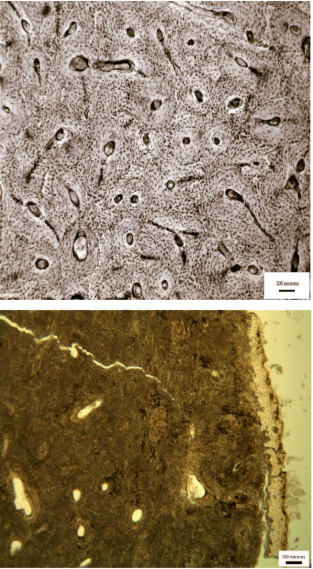
The answer may lie in geochemical techniques. Bioarchaeologists have long-recognised that tissue chemistry can give insight into childhood experience. Changes to isotopic ratios in bones, for example, can help to pinpoint when weaning was occurring. More recently though incremental isotopic techniques have been developed that allow us to look at experiences over the life course – not just at a single point in time. Tissues that grow at known rates (like teeth, hair and nails) can be sliced into increments. Each of these increments represents a period of time in a person’s life and we can use incremental values to build a profile, showing changes to tissue chemistry over time. Why is that a big deal? Because it means we can look at changes to tissue chemistry in archaeological infants and children to see changes to diet and physiological stress over time, leading up to time of death. But perhaps more importantly we can look at the early-forming tissues of adults who survived childhood, to get an insight into their childhood experience and whether or not it was different to our non-survivors. Goodbye osteological paradox (ok, that’s an exaggeration, but it’s a good step on the way to removing bias).

Julia Beaumont (Bradford) and Janet Montgomery (Durham) are pioneering this kind of work, showing maternal and infant stress levels in Irish famine samples, and investigating the implications these have for survival. They’ve shown that they can see differences in weaning behavior between survivors and non-survivors and evidence for maternal stress in the increments that form while the infant is still in the womb. They’ve even spotted evidence for the introduction of famine relief food in the form of ‘Indian meal’ (maize), which handily has a carbon isotope signal that is very different to the much more negative values of the traditional Irish diet. The work being done on childhood during the Irish famine is extremely cool and, because it’s a relatively well-documented historical event, there are written sources like workhouse records that researchers can use to add to and support their interpretations. Lucky them!

jonnys-records

Excerpt from the Minutes of the Board of Guardians, Kilkenny Union (9 March 1843), describing the death from starvation of an infant of 2 months in the workhouse. Photo taken by Jonny Geber at the Kilkenny County Library (Local Studies 6/2K).

Work in modern and historic contexts is building up a picture of the myriad of different isotopic changes which can occur during childhood. In these contexts we either have very good knowledge of the childhood experience (as in clinically examined infants) or can extrapolate it from historical records (as in the Irish famine context). As incremental isotopic techniques are increasingly applied we are building up what is effectively a reference library, showing which changes might be related to weaning, which might relate to the introduction of complementary foods. We can also see what maternal-infant stress transfer might look like isotopically, and identify stress spikes throughout tissue formation.

This is especially useful for people like me, who work in prehistoric contexts. Here the childhood experience is very much a mystery, and having references from which to interpret isotopic results becomes important. For my work in the Atacama Desert of northern Chile it’s particularly useful because there are so many competing influences over the isotopic composition of tissues. I need all the help I can get in my interpretations!

The Atacama Desert is a crazy place to live, and it’s a crazy place isotopically. To start with the aridity of the desert environment means that terrestrial food sources have isotopic ratios that are well outside of what we’d consider ‘normal’ elsewhere. Secondly, baseline stress levels are likely to be through the roof. The Atacama is the driest hot desert on earth (fun fact: technically Antarctica is a drier ‘desert’, because all its water is tied up in the form of ice/snow). Where I work there are snowmelt fed rivers which have allowed people to farm the valleys since around 1500BC, but even then it’s precarious. So our isotopic profiles are probably going to be affected by stress at least as much as they are affected by infant/child feeding practices. In fact, there’s a prevailing theory in the area that infants and children weren’t just under nutritional stress, they were also being systematically poisoned by the extremely high heavy metal content (especially arsenic) of the rivers. Good times.

san-jose

The San Jose ‘river’ bed, Arica. Taken by the author, November 2014. Water is a precious resource in this area, and the river is used extensively for crop irrigation, meaning by the time it reaches the sea in the city of Arica, for much of the year there is no water there at all.

As a final complicating factor, the area has played host to a variety of different polities, including the Tiwanaku people and later the Inkas, who are likely to have brought with them useful complementary foods such as maize, but also different cultural expectations regarding infant/child care and feeding. But in a prehistoric context we can’t be totally sure what these were. We have some tantilising hints from later Spanish ethnographers who observed the Inka, and occasionally wrote about their childcare practices. As per usual though, these accounts tend to focus on royalty and royal males in particular, things as ‘mundane’ as women and children rarely get a look in. The brief mentions of Inka childcare do paint a picture of a rather laissez-faire attitude to young ones, with multiple accounts speaking of how it was considered weakness to hold babies, and Garcillaso de la Vega talking of keeping infants in holes in the ground beyond a certain age. There’s even a potentially (hopefully?!) exaggerated mention of sending them to work in the silver mines for misbehavior. Not very useful on the whole though, and in the northern Atacama, which was on the periphery of the Empire, these customs may not even have applied.

chicha-vessel

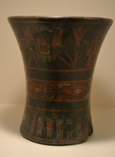
Vessels like this kero (qero) appear in the archaeological record from the Middle Horizon (450-900AD) onwards. Used for the ceremonial drinking of chicha (maize beer), they highlight the incoming of external polities and their customs. Photo is of a Tiwanaku period kero in the collections of the Museo Larco, Lima and was taken by the author.

poma-child-in-crib

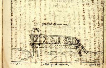
Illustration of a swaddled Inka infant in their crib from Guaman Poma’s ethnography “nueva corónica y buen gobierno”. Accessed online through Det Kongelige biobiotek

All of these things combine to make a gloriously chaotic picture of early life in the Atacama. In looking at incremental isotopic profiles from my individuals, we have evidence for almost every kind of infant/child life-experience you can imagine. Some show broadly what we’d expect for a child in any context – a signal for breastfeeding, followed by a gradual shift down to adult isotopic ratios as weaning occurs. Other profiles are dominated by stress signals, with high maternal nitrogen isotope ratios probably signifying maternal stress, and continued stress throughout infancy. We can see the use of maize as a complementary food during weaning for some individuals, but others from the same time period seem to completely ignore it, weaning onto different resources instead.

cartoon-neater

Isotopic systems can be complicated. Particularly when you work in a desert.

All this isotopic chaos can be pretty frustrating. There was no uniformity in weaning behavior in any time period in the Atacama. No massive changes with the incoming of agriculture. No population-wide processes apparent at all. How am I supposed to get my high-impact, world-changing publications now?

But actually these incremental techniques we are begin to reveal more and more complexity in decision-making, and diversity in life experiences in the past. There is no single story of childhood in the Atacama, just like there was no single story during the Irish Famine, and no two childhoods are the same today. Using these new methods we’re building individual profiles, not population models. We can see more detail so of course the picture is going to get more complicated. And in many ways that’s what bioarchaeology is about – seeing the complexity of life, and giving the people whose remains we study back their own, individual stories.

Guest post written by Dr Charlotte King, Rutherford Postdoctoral Fellow at the University of Otago (@showmethemummy) – bioarchaeologist, traveller and adventure-hunter. Big fan of isotopic systems, and desperately searching for agricultural origins.
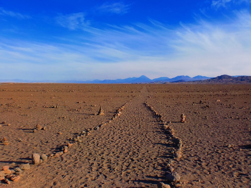
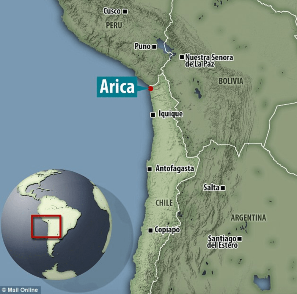
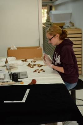


Take a sneak peek at our new resource on the “Bioarchaeology of Childhood” coming soon to Oxford Bibliographies in Childhood Studies

We have a forthcoming large annotated bibliography on the Bioarchaeology of Childhood coming soon to Oxford Bibliographies online. Take a sneak peek here. This will be useful to all bioarchaeology and human osteoarchaeology students, and academics for research and teaching. Please contact me here to request a personal copy.

Note that this is now published online

Halcrow, Siân E.; Ward, Stacey M. “Bioarchaeology of Childhood.” In Oxford Bibliographies in Childhood Studies. Ed. Heather Montgomery. New York: Oxford University Press, forthcoming.

fig.3asia2000report

To Achieve the Impossible: Research and study leave with children

Recently, there has been a study published by researchers at my own University on the experience of Research and Study Leave (RSL) or sabbatical for men and women. It found  that families are negatively affected to taking RSL with international travel due to childcare requirements and associated costs.

I am lucky that I am in a permanent position and at a University that supports RSL. I am also ‘lucky’ that I have recently sold my house. The small proceeds from this have allowed me to pay for my 2- and 11-year-olds airfares and childcare, which has thus far cost over NZ$15,000, plus continued payment of daycare fees to keep the enrollment of my 2-year-old at our University childcare.

What I am truly lucky for is the child-centered cultures that I work in and the amazing colleagues and students I have who accommodate them. The best place in accommodating my children has been in Thailand and Laos where friends and my local nanny have been absolutely fabulous. I have tried to plan this stint of fieldwork so as my 11-year-old is away at a time that includes her school break and to work around a visiting fellowship to the UK at the end of the year. However, this timing has also meant that it is HOT and hard for my kids. My 11-year-old misses her friends, but she has been extraordinarily self-motivated at doing her schoolwork each day (even in the weekends) working on her maths, reading and writing. I actually have to tell her to stop doing it at times so she gets out of the house!

Research highlights thus far have been working on the human remains from the Plain of Jars site in Laos excavated under the direction of Dougald O’Reilly and Louise Shewan. This site is under consideration for World Heritage Status and has gained archaeological interest from researchers around the world. I have also been continuing with my data collection from the infants and children from a Thai Iron Age site (see my post from early this year). This season I have found several very pre-term infants. This is of significance in indicating poor maternal health in this past population, and further supports our developing model of health change during this turbulent time of agricultural and social change.

IMG_3259
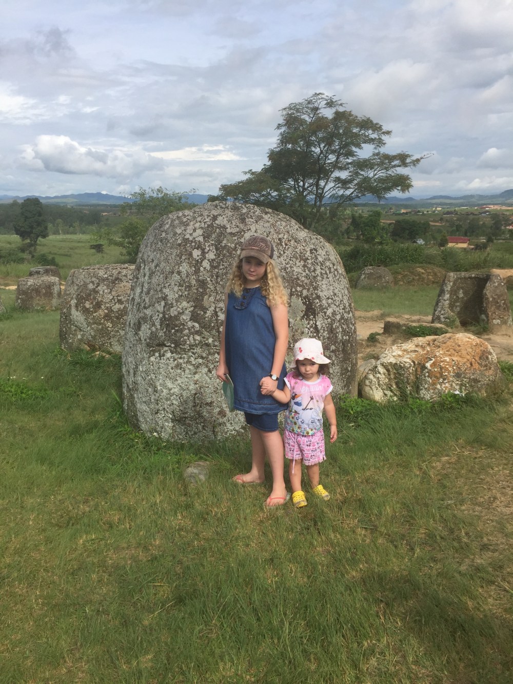
Our visit to the Plain of Jars site 1.


IMG_2969A 24-26 week old foetus from the Iron Age site of Non Ban Jak, Northeast Thailand.
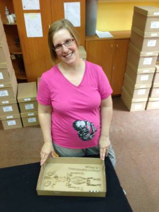
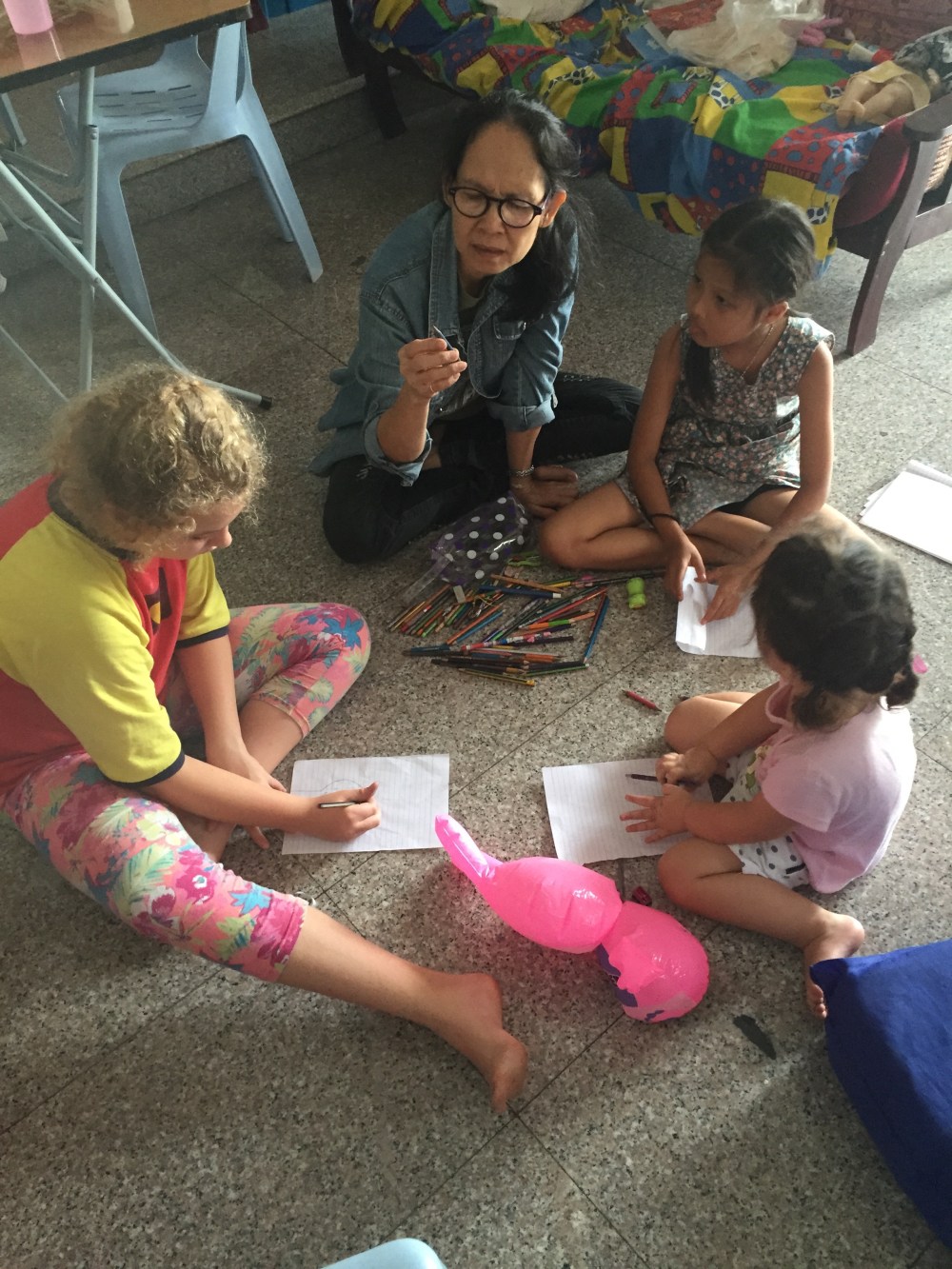
 

IMG_2978Our “super-nanny”.

The most difficult place we have been this year for accommodating children was the US for two major conferences. Childcare was US$200 a day plus extra expenses. Neither of the conferences provided childcare services, which I would have been very happy to pay for. Thank goodness for two local moms at the first conference who traveled to the store to buy us some groceries while we were stuck in a food desert! Despite the expense, both conferences have been extremely beneficial for my research. I have established new collaborations, been invited to visit universities, and they were invaluable for me to keep up-to-date with recent research developments in my field. I was also able to support two of my students who attended the conferences.

I’m happy that my RSL so far has been possible with my children. Without the ability for international travel I can’t do my research or attend major conferences. However, next time I will try to be more realistic about my plans with the kids. They are enjoying their time in Southeast Asia but the logistics and financial issues are a lot of pressure.

We are off to the UK in September until December for my fellowship to work with colleagues in the Department of Archaeology at the University of Durham. Another place with supportive colleagues! I’m looking forward to the next adventure!

IMG_3315The two-year-old helping me re-box some archeological human remains.

IMG_2971.jpg
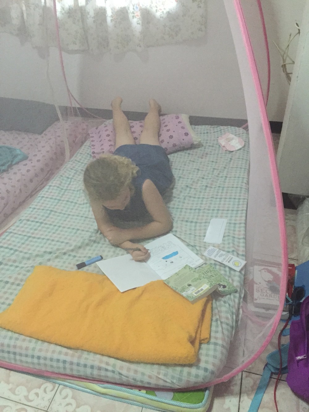
The 11-year-old hiding in our bedroom for some quiet space to do her school work under the mosquito net.
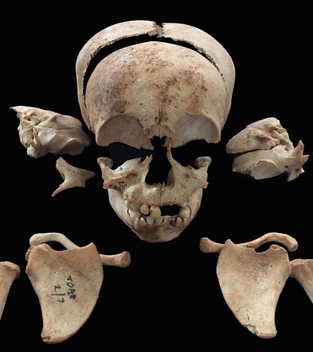
 

Is this really a 5,000 year old mother and baby?

A recent story of a 4,800-year-old ‘mother’ cradling a baby has been pulling at the heart strings of people worldwide with sensationalist headlines such as “Mother’s enduring love for baby revealed as 5000-year-old fossil found” and “Fossil of 5000-year-old mother cradling baby found in Taiwan”. But is this story everything it’s really cracked up to be?

An archeological team working at a Neolithic site near the city of Taichung since 2014 has unearthed “48 sets of remains”, presumably the number of individual graves, representing the earliest burial site in Taiwan. One of these burials has been described as a mother and baby. However, the news accounts provide little information as to why the researchers believe this to be the case, apart from the placement of the baby with the adult female and the turning of her head to be “looking at her baby” (Figure 1).

Screen Shot 2016-04-29 at 4.44.59 pm
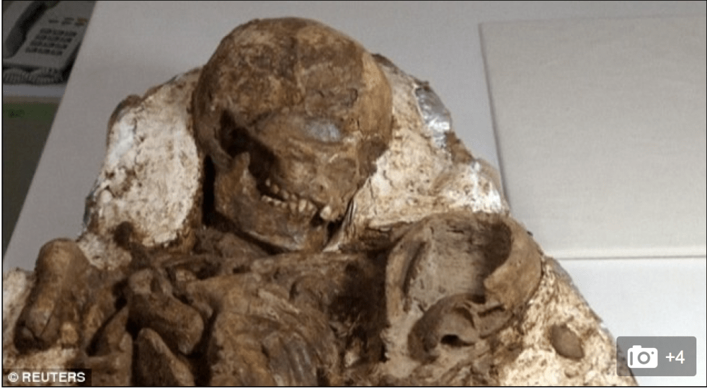
Figure 1: The 4800-year-old “mother and baby” found in Taiwan (source: Reuters)

It is likely that if a female and newborn baby is found in a burial context that they died during childbirth (see my earlier post on fetuses in archaeology). Childbirth is the most critical time for both a mother and baby. This has even led some archaeologists to argue that higher mortality rates of young adult females compared with males represent the hazards of childbirth in the past.

The baby has been described as a foot and a half (about 46 cms), which is about the size of a newborn baby. However, looking at the photos and the videos from the news stories the baby looks too big to be a newborn. The only bones present seem to be from the waist-up. Looking at the relative size of the hands of the archaeologist cleaning the bones and the upper body of the baby (Figure 2), it may be that the size cited is for the upper body, supporting that the infant is older than a newborn. It is difficult to see the cranial bones to assess their development to infer an age-at-death. The cranial bones look thicker than a newborn, but it is unclear as it appears there is some concreted soil adhering to the surface of the bones. Given that this infant seems older than a newborn it is unlikely that they were mother and child.

Screen Shot 2016-04-29 at 2.47.40 pmFigure 2: Archaeologist cleaning the ‘mother-baby’ burial (photo: Reuters video).

In a small Neolithic community there may have been some kind of relationship between the adult female and the infant, or they may have only been buried together because their deaths coincided. Using a cross cultural example, in the Anglican burial tradition babies were interred with non-maternal women in instances of coinciding death (Roberts and Cox 2003: 253).

To assess if there is a biological relationship between this purported mother-baby pair, ancient DNA analyses could be undertaken, but this is difficult with preservation issues in tropical contexts. We should also keep in mind that a mother-child relationship is not always biological.

The fact that the adult female had her head turned to her left may be the result of the burial environment, as some bones can shift in open spaces such as coffins, or from the weight of soil on the bones. Further research looking at the positions of the bone could give more insight on the mode of burial.

We will have to await the scientific presentation of the findings from this site to evaluate the likelihood for this purported mother and baby.

Bacterial bioerosion of bone may help identify stillborn infants from the past

New research using novel microscopic investigation of bacterial bioerosion of archaeological bone has shown that you can differentiate between stillborn and post-newborn babies. This was most exciting to me as offering a means to contribute to the debate of the interpretation of infanticide in the past, through an investigation of time of death.

Bioerosion is the removal of mineralised substrate through the action of organisms, and has been found to be the most common form of microbial attack of archaeological bone (Figure 1). The author of this new research, Tom Booth from the Natural History Museum, notes that although it was once believed that soil bacteria caused most of this bioerosion in bone, it is the gut microbia that is responsible for corpse putrification that causes this process. Based on the findings that it is the bacteria inside the body that produces this bioerosion, the author thought that this could be useful for assessing different mortuary treatments of the body.

Screen Shot 2016-03-12 at 9.06.06 pm

Figure 1: Transmitted light micrograph of a human fresh bone transverse femoral thin section (top) demonstrating perfect microstructural preservation and a typical archaeological femoral section (bottom) where the internal microstructure has been extensively altered by bacteria (from Booth et al., 2015).

To investigate if there is any relationship between bacterial bone bioerosion and funerary treatment, Booth undertook a microscopic analysis of human bones from European prehistoric (4000 B.C. – A.D. 43) and British historical (A.D. 43 – present day) sites. These two assemblages were used as they have been found to have different funerary practices, with the historic period sites practicing burial soon after death, whereas the prehistoric sites have more variable mortuary practices, sometimes including postmortem modification. E.g. Booth and colleagues’ work that found evidence for mummification in Bronze Age Britain using this microscopic method has recently received media attention.

This research shows that irrespective of burial environment, including antiquity or soil type, there was immaculate histological preservation of almost half of the neonatal samples. This is interpreted as the result of sterility of stillborn infant intestinal tracts resulting in the bones being unaffected by the process of bacterial tunneling. In addition, most (12/15) of the unbioeroded newborn samples are from historical cemeteries where most of the other samples had been extensively bioeroded. A previous experimental study by White and Booth using pigs found that bone from stillborn neonatal carcasses had immaculate histological preservation due to the intrinsic sterility of newborn infant intestinal tracts.

Booth found that the soil type had no relationship with bacterial bioerosion. There was evidence for variation in bacterial bioerosion among the later prehistoric assemblages argued to be “consistent with the knowledge that these individuals were subject to variable early post mortem treatment that exposed the bones to diverse levels of bacterial attack.” Bacterial bioerosion in the historical assemblage was high, consistent with that expected within bones of intact bodies that had been interred soon after death.

The use of this novel method to differentiate stillborn vs post-newborn infants can contribute to extending our knowledge of the cause of death during the most crucial time for mother and child in the past, and may also have useful applications for the study of cultural beliefs around stillbirth and post-neonatal death.

References:

Booth, T. J., A. T. Chamberlain and M. P. Pearson (2015). “Mummification in Bronze Age Britain.” Antiquity 89(347): 1155-1173.

Fetuses in bioarchaeology

The concept of fetuses in archeology probably brings to mind poignant images of the tiny bones of a baby in the pelvic cavity of a female adult skeleton, although finds such as these are actually rather rare. In practice, many bioarchaeologists apply the description of ‘fetus’ to babies from bioarchaeological samples identified as younger than 37 weeks gestational age (e.g. Halcrow et al. 2008; Lewis and Gowland 2007; Mays 2003; Owsley and Jantz 1985). However, there are problems associated with estimation of age-at-death of these babies, who may indeed be fetuses, but also may be premature births, or small-for-gestational age full-term births. If the medical definition of a fetus as an unborn baby is applied (Forfar et al. 2003; Halcrow and Tayles 2008; Lewis and Gowland 2007; Scheuer and Black 2000), the in-utero skeletons would seem to represent the only finds in archaeology that can be confidently identified as fetuses. However, even an apparent in-utero fetus may in fact have been a neonate mortality, illustrating the care with which research in this field needs to be completed.

Generally little bioarchaeological research considers fetuses. For example, some growth studies and demographic analyses do not include preterm infants because of lack of comparative fetal bone size data (e.g. Johnston 1961). Also, the attention afforded to purported evidence of infanticide, based primarily on the reported high number of perinates in some skeletal assemblages (see my previous blog story on this), has deflected interest away from the contributions that fetuses can make to understanding bioarchaeological questions, including maternal health and disease and social organization from mortuary ritual analyses (Bonsall 2013; Faerman et al. 1998; Gilmore and Halcrow 2014; Mays and Eyers 2011; Mays 1993; Mays and Faerman 2001; Smith and Kahila 1992).

It is believed that approximately 3 in 10 pregnancies are spontaneously aborted, with the majority of these occurring in the first trimester, most being the result of genetic abnormalities (Fisher 1951). First trimester fetuses are very unlikely to be recovered in the bioarchaeological context. Bone development does not start until approximately six–eight weeks gestational age, and any bone formation prior to the second trimester would be unlikely to be preserved because of the low level of mineralization, and/or would be extremely difficult to identify in an archaeological context. The only first trimester fetus reported from an archaeological context is from the Libben sample, Ohio, a Late Woodlands site occupied 8th-11th century AD (White 2000: 20, see figure 1). There are published instances of preserved fetal individuals from the second trimester, e.g. the well-preserved fetus of 20 weeks gestational age from the Kellis 2 site, Dakhleh Oasis, Egypt (Wheeler 2012: 223). Owsley and Jantz (1985) have found three fetuses younger than 28 weeks gestation at Arikara sites in South Dakota. Hillson (2009) has also reported the findings of fetuses as young as 24 gestational weeks from a large Classical period infant cemetery at Kylindra on Astypalaia, in Greece.

Screen Shot 2016-02-06 at 5.37.32 pm

Figure 1. Fetal skeletal material from the prehistoric Libben site, the smallest burial ever recorded (from White et al. 2011: 329). The long bones measure less than 2 cms.


Types of fetus burials

Differentiating burial types has the potential to contribute to research on maternal health, and the cause of death for the mother and child in the past. For example, a premature birth is more likely to indicate poor health and/or nutritional status of a woman, compared with a baby who died around full-term from obstructed labor. Distinguishing the type of fetal death and burial, whether the baby was full-term, or a pre-term or small-for gestational age baby, in conjunction with evidence of stress and diet and of both the mother and baby may give insights into overall health in past populations (Figure 2).


Halcrow Fig. 8 copyFigure 2. Infant jar burials from the Iron Age site of Noen U-Loke, NE Thailand. Left: full-term infant, approximately 40 gestational weeks (burial 100); right: pre-term infant, or ‘fetus’, approximately 30 gestational weeks (burial 89). (Photograph courtesy of C.F.W. Higham)

In-utero fetuses

If the skeletal remains of a baby are found crouched in a fetal position within the pelvic cavity of an adult female, the mother likely died while the fetus was in-utero, before or during labor. The pregnant woman may therefore have died due to pregnancy or labor complications (Lewis 2007: 34). There is very little evidence for in-utero fetuses in the bioarchaeological context. Approximately 20 cases of pregnant or laboring females (i.e., interred with fetal remains in-situ) have been published in the archaeological literature, being argued to represent complications from childbirth (e.g. Ashworth et al. 1976; Cruz and Codinha 2010; Hawkes and Wells 1975; Högberg et al. 1987; Smith and Wood-Jones 1910, in Lewis 2007; Lieverse et al. 2015; Malgosa et al. 2004; O’Donovan and Geber 2010; Owsley and Bradtmiller 1983; Persson and Persson 1984; Pounder et al. 1983; Rascon Perez et al. 2007; Sjovold et al. 1974; Roberts and Cox 2003; Wells 1978).

The dearth of literature on in-utero fetuses in bioarchaeology may not be due to absence of evidence, but rather from the small bones being missed or misidentified during excavation, or reported only in the grey literature. There are numerous accounts of fetuses being misidentified as animal bones during excavation (e.g. Ingvarsson-Sundström 2003). For example, Roberts and Cox (2003) have reported at least 24 unpublished cases of fetuses from British excavations. There are further instances of fetal bones being found co-mingled with adult burials post-excavation, which may represent a baby in-utero, or a possible mother and baby post-birth burial (S. Clough, pers. comm.).

Bioarchaeologists have reported on cases of purported obstructed labor causing maternal and fetal perinatal death based on positioning of the fetus in the pelvic cavity or the finding of preterm mummified remains in-utero (Arriaza et al. 1988; Ashworth et al. 1976; Lieverse et al. 2015; Luibel 1981; Malgosa et al. 2004; Wells 1975).

Post-birth ‘fetuses’

If a perinate is found buried alongside an adult, with the same head orientation, then the infant has been buried post-birth, whether naturally or by caesarian section (Lewis 2007: 34) (Figure 3). In some contexts it is very common for newborns to be placed on the chest of adult women (presumably their mother) (Standen et al. 2014). To identify post-birth ‘fetuses’ archaeologically, if the majority of the infant remains are in the pelvic cavity of the adult, yet the legs are extended and/or the cranium lies among the ribcage, then the baby may have been delivered and then placed on top of the mother’s (or other adult’s) torso during burial (Lewis 2007: 34). It is argued that as both mother and baby bodies’ skeletonize, the baby’s bones can become settled among the mother’s ribs and vertebrae. This is important to note as these neonates may be mistaken for breech, obstructed labors in the archaeological context (e.g. Willis and Oxenham 2013). Willis and Oxenham (2013) describe an ‘in-utero breech’ presentation of a 38 gestational week fetus from Neolithic Southern Viet Nam. They describe the cranium as “below the mothers right lower ribs” (it is not clear if they mean inside the abdominal/thoracic cavity or inferior to the right lower ribs) and the postcranial skeleton as “extended down toward the mothers pelvis” with the left femur “positioned within the mothers pelvic cavity and a tibia … positioned beside [lateral] the lesser trochanter of the mothers right femur.” They also state the “right pars lateralis [part of the base of the occipital bone of the cranium] was concreted to the anterosuperior portion of the shaft of the 10th right rib of the mother, near the sternal end.” Given this partially extended (non-fetal) positioning and the part of the cranial base being found anterior to the rib cage), it could be possible that the baby was not in the abdominal cavity, but placed on top of the mother’s torso after birth.

IMG_0879

Figure 3. Full-term neonate (burial 48) buried alongside an adult female (burial 47) from Khok Phanom Di (photograph courtesy of C.F.W. Higham). This could possibly represent a perinate and mother who died from complications during or following childbirth.

Ancient DNA analyses may be used to assess the relationship of the adult and fetal burials where the fetus has been placed on the purported mother, or the archaeological context is unclear. Lewis (2007: 35) has argued that this is important to distinguish these relationships, as in some contexts, e.g. in the Anglican burial tradition, babies were interred with non-maternal women in instances of coinciding death (Roberts and Cox 2003: 253).

Multiple fetal pregnancies and births

There have been two reported instances of twin fetuses in-utero in the bioarchaeological literature (Lieverse et al. 2015; Owsley and Bradtmiller 1983), with others found in a post-birth context. There has been a recent increase in the interest in multiple births in bioarchaeology, including an investigation of social identity and concepts of personhood through the investigation of mortuary treatment (e.g. Einwögerer et al. 2006; Halcrow et al. 2012). Human twins are rare, with approximately one occurrence for every 100 births (Ball and Hill 1996). However, they appear in the literature more commonly than expected, compared with singleton fetuses (e.g. Black 1967; Chamberlain 2001; Crespo et al. 2011; Einwögerer et al. 2006; Flohr 2014; Halcrow et al. 2012; Lieverse et al. 2015; Owsley and Bradtmiller 1983). This is probably because they are seen as more significant by the archaeologist.

An example of a possible twin burial was found in an Upper Paleolithic site of Krems-Wachtberg, Austria (Einwögerer et al. 2006). The infants from this double burial were identified as twins from their identical age (as estimated from their dentition), same femora size and their simultaneous interment (both estimated at full-term age at death). Interestingly the bodies lay under a mammoth scapula and a part of a tusk and were associated with 30 ivory beads. Einwögerer et al. (2006) suggest, based on this mortuary evidence, that these newborns were an important part of their community. Another case of a twin burial is from the mid fourth-century site of Olèrdola in Barcelona, Spain (Crespo et al. 2011). The two newborns were found at the same stratigraphic level with their lower limbs entwined, indicating that they were buried simultaneously. We (Halcrow et al. 2012) havev also presented an extremely rare finding of at least two and possibly four twin burials from a 4,000-3,000 year old BP Southeast Thailand site, offering a methodological approach for the identification of archaeological twin (or other multiple birth) burials and a social theoretical framework to interpret these in the past.

Post-mortem birth (‘coffin-birth’)

Post-mortem birth or ‘coffin-birth’ refers to the occurrence of fetuses that were in-utero when the mother died and were expelled after burial (O’Donovan and Geber 2010) (Figure 4). This is also talked about by Katy Meyers Emery in her blog story on coffin birth in her blog Bones Don’t Lie. Post-mortem birth by fetal extrusion has been documented in rare forensic cases from the build up of gas within the abdominal cavity resulting in the emission of the fetus (Lasso et al. 2009; Schultz et al. 2005). Lewis (2007: 34-37, 91) and O’Donovan et al. (2009) argue that if fetal remains are complete and in a position inferior to and in-line with the pelvis outlet, with the head oriented in the opposite direction to the mother, then there is the possibility of coffin birth (Figure 3). If they lie within the pelvic outlet, this means that there was partial extrusion during decomposition (Hawkes and Wells 1972). However, partial extrusion could also be the result of an obstructed labor of a baby in the breech position, but this would likely result in extrusion of the lower limbs. Sayer and Dickenson (2015) argue that postmortem fetal extrusion is implausible under some burial conditions and with that decomposition of the baby in-utero would mean that it isn’t likely to be birthed from an undilated cervical canal. This, however, assumes that there was no dilation at the time of death of the mother.

3

Figure 4. Potential coffin birth (from Appleby et al. 2014)

Social identity

The investigation of mortuary treatment of pregnant women may give us information on social identity related to childbearing and fetuses themselves. For example the discovery of a 34-36 week old fetus cremated with the ca. 850 B.C. “Rich Athenian Lady” led to a recognition that her grave wealth may have been related to her dying while pregnant or during childbirth, rather than primarily her social status (Liston and Papadopoulos 2004).

Research of the archaeology of grief is starting to consider community members’ responses to infant and fetal death (e.g. Cannon and Cook 2015; Murphy 2011). The purported marginalization of fetuses along with infants in the archaeological record, including location and simplified mortuary treatment has led some scholars to interpret that they were of little concern beyond immediate family members (Cannon and Cook 2015). Considering literature on intense grief after miscarriage and infant death starts to challenge the notion that their loss was of little consequence (Murphy 2011).

NB: Part of this story is from the chapter:

Halcrow, S.E., N. Tayles and G.E. Elliott (2016 expected) The Bioarchaeology of Fetuses. In Han S, Betsinger TK, and Scott AB; The Fetus: Biology, Culture, and Society. Berghahn Books. (under contract)

References

Appleby J, Seetah TK, Calaon D, Čaval S, Pluskowski A, Lafleur JF, Janoo A, and Teelock V (2014). ‘The Non-Adult Cohort from Le Morne Cemetery, Mauritius: A Snap Shot of Early Life and Death after Abolition.’ Int J Osteo 24(6): 737-746.

Arriaza B, Allison M, and Gerszten E. 1988. ‘Maternal Mortality in Pre-Columbian Indians of Arica, Chile’. Am J Phys Anthropol 77:35-41.

Ashworth JT, Allison MJ, Gerszten E, and Pezzia A. 1976. ‘The Pubic Scars of Gestation and Parturition in a Group of Pre‐Columbian and Colonial Peruvian Mummies’. Am J Phys Anthropol 45(1):85-89.

Ball HL, and Hill CM. 1996. ‘Reevaluating “Twin Infanticide”‘. Curr Anthropol 37(5):856-863.

Black GA. 1967. Angel Site: An Archaeological, Historical, and Ethnological Study. Indiana Historical Society: Indianapolis.

Bonsall L. 2013. ‘Infanticide in Roman Britain: A Critical Review of the Osteological Evidence’. Childhood in the Past 6(2):73-88.

Cannon A, and Cook K. 2015. ‘Infant Death and the Archaeology of Grief’. Cambridge Archaeological Journal 25(2):399-416.

Chamberlain G. 2001. ‘Two Babies That Could Have Changed World History’. Historian 72:6-10.

Crespo L, Subira ME, and Ruiz J. 2011. ‘Twins in Prehistory: The Case from Olerdola (Barcelona, Spain; 2. IV II BC)’. Int J Osteo 21:751-756.

Cruz C, and Codinha S. 2010. ‘Death of Mother and Child Due to Dystocia in 19th Century Portugal’. Int J Osteo 20(4):491-496.

Einwögerer T, Friesinger H, Händel M, Neugebauer-Maresch C, Simon U, and Teschler-Nicola M. 2006. ‘Upper Palaeolithic Infant Burials’. Nature 444(7117):285.

Faerman M, Kahila Bar-Gal G, Filon D, Stager L, Oppenheim A, and Smith P. 1998. ‘Determining the Sex of Infanticide Victims from the Late Roman Era Through Ancient DNA Analysis’. J Archaeol Sci 25:861-865.

Fisher RS. 1951. ‘Criminal Abortion’. J Crim L Criminology & Police Sci 42:242.

Flohr S. 2014. ‘Twin Burials in Prehistory: A Possible Case From the Iron Age of Germany’. Int J Osteo 24:116-122.

Forfar JO, Arneil GC, McIntosh N, Helms PJ, and Smyth RL, editors. 2003. Forfar and Arneil’s Textbook of Pediatrics. 6th ed. Edinburgh: Churchill Livingstone.

Gilmore HF, and Halcrow SE. 2014. “Sense or Sensationalism? Approaches to Explaining High Perinatal Mortality in the Past”. In: Thompson JL, editor. Tracing Childhood: Bioarchaeological Investigations of Early Lives in Antiquity. Florida: University of Florida Press. p 123-138.

Halcrow SE, Tayles N, Inglis R, and Higham C. 2012. ‘Newborn Twins from Prehistoric Mainland Southeast Asia: Birth, Death and Personhood’. Antiquity 86:838-852.

Halcrow SE, and Tayles N. 2008. ‘The Bioarchaeological Investigation of Childhood and Social Age: Problems and Prospects’. Journal of Archaeological Method and Theory 15(2):190-215.

Halcrow SE, Tayles N, and Livingstone V. 2008. ‘Infant Death in Prehistoric Mainland Southeast Asia’. Asian Persp 47:371-404.

Hawkes SC, and Wells C. 1975. ‘Crime and Punishment in an Anglo-Saxon Cemetery?’. Antiquity 49(194):118-122.

Hillson S. 2009. ‘The World’s Largest Infant Cemetery and its Potential For Studying Growth and Development’. Hesperia Supplement:137-154.

Högberg U, Iregren E, Siven C-H, and Diener L. 1987. ‘Maternal Deaths in Medieval Sweden: an Osteological and Life Table Analysis’. Journal of Biosocial Science 19(4):495-503.

Ingvarsson-Sundström A. 2003. Children Lost and Found: A Bioarchaeological Study of the Middle Helladic Children in Asine With a Comparison to Lerna [PhD thesis]. Uppsala: Uppsala University.

Johnston FE. 1961. ‘Sequence of Epiphyseal Union in a Prehistoric Kentucky Population from Indian Knoll’. Hum Biol 33:66-81.

Lasso E, Santos M, Rico A, Pachar JV, and Lucena J. 2009. ‘Expulsión Fetal Postmortem’. Cuadernos de Medicina Forense (in Spanish) 15: 77–81.

—. 2007. The Bioarchaeology of Children: Perspectives from Biological and Forensic Anthropology. Cambridge: Cambridge University Press.

Lewis ME, and Gowland RL. 2007. ‘Brief and precarious lives: Infant Mortality in Contrasting Sites From Medieval and Post-medieval England (AD 850-1859)’. Am J Phys Anthropol 134:117-129.

Lieverse AR, Bazaliiskii VI, and Weber AW. 2015. ‘Death by Twins: A Remarkable Case of Dystocic Childbirth in Early Neolithic Siberia’. Antiquity 89(343):23-38.

Liston MA, and Papadopoulos JK. 2004. ‘The” Rich Athenian Lady” Was Pregnant: The Anthropology of a Geometric Tomb Reconsidered’. Hesperia:7-38.

Luibel AM. 1981. Use of Computer Assisted Tomography in the Study of a Female Mummy from Chihuahua, Mexico. Annual Meeting of the Southwestern Anthropological Associatation. Santa Barbara.

Malgosa A, Alesan A, Safont S, Ballbe M, and Ayala MM. 2004. ‘A Dystocic Childbirth in the Spanish Bronze Age’. Int J Osteo 14(2):98-103.

Mays S. 1993. ‘Infanticide in Roman Britain’. Antiquity 67:883-888.

Mays SA, and Faerman M. 2001. ‘Sex Identification in Some Putative Infanticide Victims from Roman Britain Using Ancient DNA’. J Archaeol Sci 28:555-559.

Mays S, and Eyers J. 2011. ‘Perinatal Infant Death at the Roman VIlla Site at Hambleden, Buckinghamshire, England’. J Archaeol Sci 38(8):1931-1938.

Murphy EM. 2011. “Parenting, Child Loss and the Cilliní of Post-Medieval Ireland”. In: Lally M, and Moore A, editors. (Re)thinking the Little Ancestor: New Perspectives on the Archaeology of Infancy and Childhood BAR International Series 2271. Oxford: British Archaeological Reports. p 63-74.

O’Donovan E, Geber J, and Baker C. 2009. ‘Archaeological excavations on Mount Gamble Hill: stories from the first Christians in Swords’. Axes, Warriors and Windmills: Recent archaeological discoveries in North Fingal. Fingal County Council.

O’Donovan E, and Geber J. 2010. “Archaeological Excavations on Mount Gamble Hill, Swords, Co. Dublin”. In: Corlett C, and Potterton M, editors. Death and Burial in Early Medieval Ireland in Light of Recent Excavations. Dublin: Wordwell Ltd. p 64-74227-74238.

Owsley DW, and Bradtmiller B. 1983. ‘Mortality of Pregnant Females in Arikara villages: Osteological Evidence’. Am J Phys Anthropol 74:331-336.

Owsley DW, and Jantz RL. 1985. ‘Long Bone Lengths and Gestational Age Distributions of Post-

Persson O, and Persson E. 1984. Anthropological Report on the Mesolithic Graves from Skateholm, Southern Sweden. I: Excavation Seasons 1980-82. Lund: University of Lund.

Pounder DJ, Prokopec M, and Pretty GL. 1983. ‘A Probable Case of Euthanasia Amongst Prehistoric Aborigines at Roonka, South Australia’. Forensic Science International 23(2-3):99-108.

Rascon Perez J, Cambra-Moo O, and Gonzalez M. 2007. ‘A Multidisciplinary Approach Reveals an Extraordinary Double Inhumation in the Osteoarchaeological Record’. Journal of Taphonomy 5:91-101.

Roberts C, and Cox M. 2003. Health and Disease in Britain: From Prehistory to the Present Day. Thrupp: Sutton Publishing Limited.

Sayer D, and Dickinson SD. 2013. ‘Reconsidering Obstetric Death and Female Fertility in Anglo-Saxon England’ World Archaeol 45(2): 285-297.

Scheuer JL, and Black S. 2000. Developmental Juvenile Osteology. San Diego, California: Academic Press.

Schulz F, Püschel K, and Tsokos, M. 2005. ‘Postmortem fetal extrusion in a case of maternal heroin intoxication.’ Forensic Science, Medicine, and Pathology 1(4): 273–276.

Sjovold T, Swedborg I, and Diener I. 1974. ‘A Pregnant Woman From the Middle Ages with Exostosis Multiplex’. Ossa 1:3-23.

Smith P, and Kahila G. 1992. ‘Identification of Infanticide in Archaeological Sites: a Case Study From the Late Roman-Early Byzante Periods at Ashkelon, Israel’. J Archaeol Sci 19(6):667-675.

Standen VG, Arriaza BT,and Santoro CM. 2014. “Chinchorro mortu­ary practices on infants: Northern Chile ar­chaic period (BP 7000–3600)”. In: Thompson JL, Alfonso-Durruty MA, and Crandall JJ, editors. Tracing Childhood: Bioarchaeological Investigations of Early Lives in Antiquity. Florida: University of Florida Press. p 58-74.

Wells C. 1975. ‘Ancient Obstetric Hazards and Female Mortality’. Bulletin of the New York Academy of Medicine 51(11):1235.

Wells C. 1978. ‘A Medieval Burial of a Pregnant Woman’. The Practitioner 221:442-444.

Wheeler SM. 2012. ‘Nutritional and Disease Stress of Juveniles from the Dakhleh Oasis, Egypt’. Int J Osteo 22(2):219-234.

White TD, Black MA, Folkens PA. 2011. Human Osteology (Third Edition). Academic Press.

Willis A, and Oxenham MF. 2013. ‘A Case of Maternal and Perinatal Death in Neolithic Southern Vietnam, c. 2100–1050 BCE’. Int J Osteo 23(6):676-684.

Infanticide in the archaeological record: sense or sensationalism? (Or: No, I’m not an ‘over-emotional’ mother and archaeologist)

A cursory look through the bioarchaeological literature for explanations of infant death in the past may leave you with a view that infants were being purposefully killed and buried in community cemeteries or simply tossed away in high numbers (e.g. Mays 1993, Mays and Eyers, 2011, Smith and Kahila, 1992).


But what is the likelihood that these accounts are accurate? Here I want to take an analytical look at the bioarchaeological evidence and arguments for infanticide. Some of my views on childhood in the past have been criticised for being clouded by my status as a mother within a ‘Western’ culture. Sometimes I feel that my interpretations are dismissed and put down to my ‘personal’, ‘irrational’, ‘hyper-emotional’, ‘ethnocentric’ thoughts on infancy. One example of this type of experience was at a conference when a senior academic after viewing my poster ‘mansplained’, “you must realise that childhood wasn’t a rosy experience like it is now where you come from. They weren’t wrapped up in cotton wool!”. I wonder if he would have said that if my infant wasn’t attending the conference, the result of no childcare options for participants (dockristy touches on this issue of inclusive conferences for caregivers in her recent blog post).


Infanticide is the intentional killing of infants. Legally, “infanticide” can refer to the deliberate killing of any infant under the age of 12 months (Kellet, 1992). Here I use the term for intentional infant killing around the time of birth, as this is the time in which it usually occurs. Infanticide has been practised in a wide range of cultures through time, and has been argued in some anthropological texts to be an adaptive strategy to environmental, economic, and social circumstances since the Pleistocene era (Hausfater and Hrdy, 1984:xxix).


Common methods for disposing of unwanted children in non-Christian cultures were exposure or drowning without subsequent burial or with covert burial (reviewed in Gilmore and Halcrow, 2014). Some of the motives documented for infanticide include poverty, and if a baby was born with a physical deformity or was “weak”. The sex of infants was also an important factor in infanticide practice for many cultures.


What evidence are these bioarchaeological studies using to inform their interpretations of infanticide? For most papers their main evidence cited for infanticide is a peak rate of mortality around the age of full-term gestation (the perinatal period of about 38-41 weeks gestation) (e.g. Mays 1993, Mays and Eyers, 2011, Smith and Kahila, 1992).*


However, we know from modern age-at-death information that it is normal to see a high rate of infant death at around full-term gestation (see Halcrow et al. 2008 for a review of this evidence). Birth is the most crucial time in a baby’s and mother’s life. Birth and the first few days of life are a dangerous time for a baby with the risk of mortality being extremely high (Kelnar et al., 1995:1). Birth complications, maternal health factors and the risk of disease are likely to have increased the incidence of perinatal deaths and stillbirths in the past. Postpartum dangers include trauma, pneumonia due to infection of the amniotic cavity (Redfern, 2007:185), and respiratory distress syndrome, particularly for pre-term or low birth-weight perinates, owing to the immaturity of the lungs. Environmental hazards for the newborn include infections, bathing in contaminated water, and tetanus due to the use of dirty instruments (Kelnar et al., 1995:6-8, Redfern, 2007:185).


Unsurprisingly, this high rate of infant death around the time of birth has also been found in the archaeological record throughout the world and during different time periods. In the majority of the prehistoric Southeast Asian sites I have worked on we find a high peak of mortality occurring around the time of birth. Other sites with this type of age distribution include Argolid in the Aegean (Angel, 1971), Roman period Britain (Mays, 1993), Southeast Europe (Boric and Stefanovic, 2004), mediaeval and post-mediaeval England (Lewis and Gowland, 2007), post-contact indigenous populations in North America (Owsley and Jantz, 1985), Roman period Egypt (Tocheri et al., 2005), and many more. Were all these cultures at these different time periods killing their infants and then burying them overtly within community cemeteries? I think not. I am not arguing that infanticide never existed in the past. However, these were often discrete events with the dead babies disposed of covertly.


IMG_0879Probable mother and newborn death from the ‘Neolithic’ site of Khok Phanom Di, Southeast Thailand. This site had a infant death representation of over 40% of the cemetery sample.

P1010598A ‘foetal’ (preterm) birth from the site of Ban Non Wat, Bronze Age, Northeast Thailand. If a live birth, this baby wouldn’t have lived for long after birth because of its immaturity.


One of these bioarchaeological papers that has interpreted the practice of infanticide is based on the Yewden Roman villa site at Hambleden in Buckinghamshire, England, which became somewhat of an archaeological “celebrity”, showcased by the BBC in 2010 (Mays and Eyers, 2011). The Hambleden site has been identified as a sophisticated “two corridor” Roman villa (Percival, 1990:531). It was first excavated in 1912 by Alfred Heneage Cocks, who reported the discovery of 103 burials, 97 of which were small infants, buried under courtyards or walls on the north side of the site (Cocks, 1921). The infant bones were recently rediscovered in a museum archive after almost a century.


Mays and Eyers (2011) have compared the perinatal age-at-death distribution pattern to other sites that have been interpreted to have an ‘infanticide’ type mortality profile. Other than that there is nothing in the mortuary or archaeological information to suggest that infanticide was probable. The burials at Hambleden are inconsistent with what is known about Roman infanticide practices. As discussed, exposure or drowning were the most usual methods employed, in which case we might expect to find infant bones as haphazard scatters in middens, remote areas of the landscape, or in wells or waterways as has been the case in Scandinavia (Wicker, 1998:215).


An understanding of the historical and ethnographic information on infanticide practices and burial, the historical or other contextual information associated with the site, infant burial practices, and mortality pattern data information is essential for assessing the likelihood for infanticide. It remains that the most parsimonious explanation for cemeteries with a peak of infant death around full-term are the result of a normal age-at-death pattern.


Why then is there a preoccupation or fascination with this idea of infanticide in the past? Were people in the past seen to be of lower moral status and therefore more likely to kill their babies? Could this continued focus on arguments of infanticide stem from an anthropological legacy of the 19th century of exploring ‘dark’, ‘primitive’ cultures, who were seen to lack intelligence and emotion?


Certainly more critical engagement with the literature on infanticide motives, practices, contextual burial information, and medical literature on the causes and timing of normal infant death offers a good approach to review evidence of infant death in the past. Even a mother with a mind ‘clouded’ by breastfeeding hormones and a ‘rosy’ view of childhood can look at the empirical evidence.

————————————————————-

*Smith and Kahila (1992) also include preterm and post-perinatal infants in their “perinate” age category. The preterm infants probably died a natural death, as is likely without modern medical intervention. In historical and modern accounts, infanticide often occurs soon after birth, so the individuals who died in the post-perinatal period were also less likely to be the victims of infanticide.


NOTE: Part of this blog post has been taken from our work in the following papers (all references cited can be found within these publications):

Gilmore, H. and S. E. Halcrow (2014). Interpretations of infanticide in the past. J. Thompson, M.P. Alfonso-Durruty and John Crandell (eds). Tracing Childhood: Bioarchaeological investigations of early lives in antiquity. Florida: University of Florida Press. 123-138.

Halcrow, S. E., N. Tayles and V. Livingstone (2008). “Infant death in prehistoric Southeast Asia” Asian Perspectives. 48 (2): 371-404.

See also Gowland et al. (2014) who offer an excellent re-evaluation of evidence for infanticide in Roman Britain.

Gowland, R. L., A. Chamberlain, & R. C. Redfern (2014). “On the brink of being: re-evaluating infanticide and infant burial in Roman Britain” Journal of Roman Archaeology Supplementary Series 96: 69-88.