SSCIP Annual Conference – University of Otago, New Zealand 2021 – Registration and conference schedule

A reminder that the 13th Annual SSCIP Conference is being held via Zoom from the 25th to 28th of October (British Summer Time). It is hosted and organised by Associate Professor Siân Halcrow of the University of Otago, New Zealand, and has been scheduled into eight short sessions over four days to accommodate the different time zones of participants.

Keynote addresses will be made by Professor Maureen Carroll of the University of York, Associate Professor Alison Behie of Australian National University, Professor Holly Dunsworth of the University of Rhode Island, and Professor Sarah Knott of Indiana University Bloomington.

If you are interested in attending any of the eight sessions you can register for free using the following link: https://www.eventbrite.com/e/society-for-the-study-of-childhood-in-the-past-conference-tickets-179748280947. After you register you will be sent a confirmation email. Zoom details for the event can be found by clicking the “View the event” button in this email.

Information regarding the conference schedule and abstracts for all the talks can be found at the following link: https://docs.google.com/document/d/1TqiAhXRo9NEiHPqTD1_OxOgh-wxf4dt0_jcIHQaWG2Y/edit?usp=sharing

Session times:

Day One / 25th October 2021 – Session One: 8am – 10am BST

Day One / 25th October 2021 – Session Two: 9pm – 10pm BST

Day Two / 26th October 2021 – Session One: 8am – 10am BST

Day Two / 26th October 2021 – Session Two: 9pm – 10pm BST

Day Three / 27th October 2021 – Session One:  8am – 10am BST

Day Three / 27th October 2021 – Session Two: 9pm – 11pm BST

Day Four / 28th October 2021, Session One: 8am – 10am BST

Day Four / 28th October 2021 – Session Two: 9pm – 11pm BST

Childhood and the development of urbanisation

The collection of papers in this special issue of Childhood in the Past edited by Francesca Fulminante showcase research on infancy and childhood with sophisticated theoretical and methodological approaches to this topic. This issue represents a significant contribution to understanding the role of children and childhood during the transition to urbanization in Europe through the lens of multiple approaches, including bioarchaeological, archaeological, cognitive developmental (palaeoanthropological), sociological and historical research on infants and children, using a variety of new analytical techniques. This issue moves chronologically from the consideration of cognitive development during prehistory to the nineteenth-century urban environment. Check it out!

Moulded terracotta, beige clay with inclusions, H 9,3 cm, W 6 cm, Veii, sanctuary of Campetti 1, Rome, Museo Nazionale etrusco di Villa Giulia, inv. n. C/168. 3rd–2nd c. BCE. See Pedrucci 2021, 236, Veii 60. Courtesy of the Museum. © Mauro Benedetti.

Online resources on infant and child bioarchaeology for teachers and students

Screen Shot 2018-02-05 at 7.53.04 PM
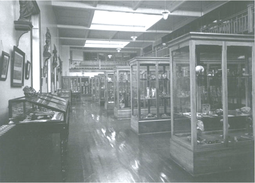
There is an increasing number of online imaging resources and software useful for the bioarchaeology of infant and children. These resources are of particular use for teaching.

Online Imaging Resources and Software

Gwen Robbins Schug from Appalachian State University (US) has developed Osteological Teaching Resources, which features a growing collection of 3D scans of human fetal and perinatal remains. Digitised Diseases, developed by the University of Bradford (UK), is a significant paleopathological resource with an impressive array of 3D models of bone pathology useful for comparative purposes. Infants and children are featured in many of their disease category types, particularly in the metabolic disease section. eSkeletons, an interactive resource for students in biological anthropology, contains limited child osteology resources. The AAOF Craniofacial Growth Legacy Collection, a database on
craniofacial development in modern children developed by the American Association of Orthodontists for clinical use, may be of use in teaching and research by bioarchaeologists. El Atlas de Osteología Infantil FACSO is being developed for teaching and learning purposes and includes infant reference material. Three-dimensional data sharing is starting to be used among paleoanthropologists for recording and sharing new fossil hominin finds, particularly because of their rare and unique nature.
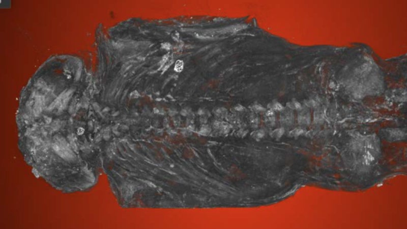
However, it is our understanding that there are no open access 3D scan data of infant and child remains. There is also little software available featuring infant and child osteology. The interactive 3D skeleton viewer Dactyl (by Anthronomics), designed by Tim Thompson from the University of Teesside (UK), has some examples of child bones. Most bioarchaeologists construct their own databases for data collection in both lab and fieldwork situations. There is an increasing number of human fetal, infant, and child casts available for purchase from Bone Clones, Inc. and France Castings for teaching and research purposes.

AAOF Craniofacial Growth Legacy Collection.

Extensive open-access database produced by the American Association of Orthodontists
Foundation (AAOF), which includes nine collections of longitudinal craniofacial growth and development records in the United States and Canada. Developed for clinicians, but also represents a useful resource for craniofacial development for teachers and researchers in the field of childhood bioarchaeology. Material includes skull, dental, and hand-wrist radiographs.

Dactyl (by Anthronomics). Tim Thompson, University of Teesside, UK.

An interactive 3D viewer loaded with photo-realistic models of bones, allowing you to rotate and zoom in on each model. The viewer comes with three preloaded adult bone elements, and there are additional packs that you can purchase, which include a “non-adult pack” with 2 bones (femur and ilium). This app is available for purchase on iTunes and can be downloaded onto an iPad.
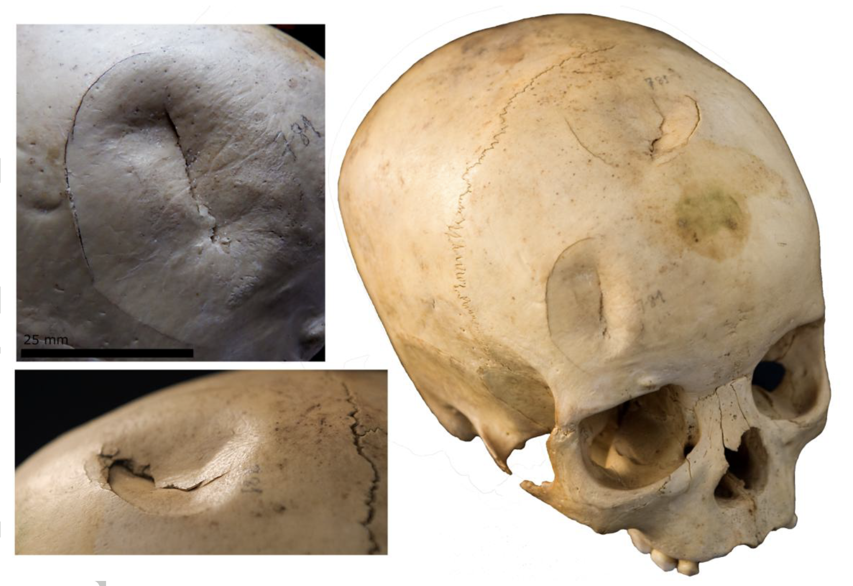
Digitised Diseases. University of Bradford, UK.

An open-access resource featuring human bones that have been digitized, with a wide range of pathological-type specimens from archaeological and historical medical collections. Infant and child pathology cases including trauma, metabolic, and infectious disease. Includes photorealistic digital representations of 3D bones that can be viewed, downloaded, and manipulated on a computer, tablet, or smartphone.

El Atlas de Osteología Infantil FACSO.

An open-access atlas project developed by students of physical anthropology at the University of Chile. This atlas is part of a larger project of developing human osteology resources for teaching and learning. The infant material is from the site of Pica 8, northern Chile, from the Late Intermediate Period.

eSkeletons. University of Texas at Austin.

Useful interactive resource to learn about human skeletal anatomy and comparative primate skeletal anatomy. Limited child osteology, including a life-size print of juvenile human bones in their teaching resources. Useful for students of all stages.
eSkeletons. University of Texas at Austin.

• Robbins Schug, Gwen. Osteological Teaching Resources. Appalachian State University.

Provides 3D scans of human fetal bones to be used for teaching purposes. The database is added to over time, with the ultimate goal to provide scans of all the skeletal elements from fetuses of different ages and perinates, cases of pathology, and traumatic injury. For access email: Robbinsgm@gmail.com

A comprehensive online resource on childhood bioarchaeology published with Oxford Bibliographies is available here free to download.

This will be useful to all bioarchaeology and human osteoarchaeology students, and academics for research and teaching.

Halcrow, Siân E.; Ward, Stacey M. “Bioarchaeology of Childhood.” In Oxford Bibliographies in Childhood Studies. Ed. Heather Montgomery. New York: Oxford University Press, 2017.

Blogs

There are a very limited number of blogs on childhood from a bioarchaeological perspective, with this blog, obviously, dedicated to the topic. The Society for the Study of Childhood in the Past has a news blog as part of their WordPress website featuring member profiles, research and conference information, and other society-related news. Other popular bioarchaeological blogs, including Powered By Osteons and, regularly include material on childhood bioarchaeology. Katharina Rebay-Salisbury has also recently started a blog called Motherhood in Prehistory, focusing on her bioarchaeological work in western Europe, which includes information on fetuses and infants in the past.

• Halcrow, Siân, and Sally Crawford. Childhood in the Past News and Blog.

This blog, established in 2015, functions to keep the members of the Society for the Study of Childhood in the Past (cited under Associations) up-to-date with news on conferences,
meetings, new research, and other opportunities in the multidisciplinary field of childhood in the past.

• Killgrove, Kristina. Powered by Osteons.

Established in 2007, this popular blog showcases Kristina Killgrove’s own research and
teaching as well as stories on recent bioarchaeological research. These blog stories often
involve child bioarchaeology. Killgrove is also a contributor for Forbes and mental_floss.

• Rebay-Salisbury, Katharina. Motherhood in Prehistory.

Established in 2015, this blog shares stories related to the author’s work investigating
motherhood in prehistoric western Europe. Often her posts include information about infants and fetuses.

Video resource on age estimation of infants and children

https://childhoodbioarchaeology.org/2019/10/16/video-on-infant-and-child-age-estimation-in-bioarchaeology/Screen Shot 2019-10-16 at 8.40.47 PM

There are numerous other educational stories on childhood bioarchaeology that will be useful for students, e.g.

Why do we have baby teeth


Skull trauma in children indicates violent pre-Hispanic Canary Island societies

There is a romanticised view that pre-Hispanic societies from the Canary Islands lived in a ‘paradise on earth’ without violence and conflict. However, recent work by anthropologists has shown that there is evidence for intentional trauma in adults from pre-Hispanic sites suggesting inter-personal violence. A recently published paper has found that the young were not spared this violence, with a high number of children from the island of Gran Canaria with skull trauma. This is significant as there is generally less evidence for violence in children compared with adults from archaeological contexts.

Screen Shot 2018-08-09 at 6.13.49 PM.png

Figure 1: A seven year old child from Guayadeque, Gran Canaria, with blunt force trauma occurring around the time of death (from Velasco-Vázquez et al. 2018).


Screen Shot 2018-08-09 at 6.46.45 PM.png

Figure 2. Areas of the skull that showed traumatic injury (from Velasco-Vázquez et al 2018).

The infants and children analysed in this research are from sites that cover an expansive part of the pre-Hispanic occupation of Gran Canaria from the 6th to 15th century CE. The authors studied a total of 65 infants and children looking to identify any sharp force trauma, puncture injuries, and blunt force trauma on the skull. Fourteen children suffered from craniofacial injuries, all of which were blunt force trauma, and two of these children have evidence that this trauma resulted in their death (e.g. Figure 1). Most of the skeletal trauma occurred on the face or forehead, a similar pattern observed in adult studies of trauma in these populations (Figure 2).

Children could have been engaged as actors in this violence as well as the victims. Although early scholars painted the Canary Islands as a peaceful and bountiful paradise, ignoring  evidence for social inequality and conflict, there is archaeological evidence for marked social hierarchy and resource depletion in this insular community, which likely led to significant social unrest.

Uncovering childhood in museums

Personal Reflections By Amanda Hoogestraat, Twitter @AmehAnthro

On my recent tour of museums in the UK, I saw small reminders of children in the exhibits featuring past societies. Children were obviously a part of every community, but are underrepresented in museum collections. There is a museum devoted to childhood in both London and Edinburgh, but perhaps other museums should consider adding more children’s items to their collections for a more balanced representation of life in the communities it displays.

For many of the museums that had childhood material culture, shoes or cradles were the only items on view.

Four out of the 55 museums that I visited had children’s skeletal remains on display; usually infants and mostly with an adult skeletons nearby. Rarely did I see older children.

However, it was the toys that interested me the most; to see how the cherished play items were very similar to those of today.

I also observed how visiting children interacted with the exhibits, especially at museums not designed specifically for them. Some of these museums had created play areas pertaining to a display nearby.

Surprisingly, the British Motor Museum was a place that had children’s programs and school tours.

I think everyone enjoys seeing items from a childhood different from our own lives or from our own childhoods. It reminds us that across time and location, children were an integral part of the society.

Egyptian ‘hawk’ mummy is a human foetus with a fatal birth defect

Recently researchers have made an unexpected discovery of a mummified foetus while CT scanning a 2300-year-old mummy known as Ta-Kush currently held at the Maidstone Museum in Kent. This coffin was labelled, “A mummified hawk with linen and cartonnage, Ptolemaic period (323 BC – 30 BC).”

Micro-CT scan shows the mummified stillborn human baby. Image: Maidstone Museum UK/Nikon Metrology UK

The high resolution CT scan results have recently been presented at the Extraordinary World Congress on Mummy Studies in the Canary Islands last month. The authors argue that the foetus was about 23-28 weeks gestation and had anencephaly as shown by underdeveloped skull bones.

To me, this begs the question as to whether the several other Egyptian ‘hawk’ mummies curated around the world are actually tiny babies. Further investigation of this baby and others will shed light on the social responses of grief and loss of those born too young to survive.

Watch here on YouTube Mummy ‘bird’ mystery

The coffin. Image: Western University


‘Freaks’ as museum exhibits: the case of the Boy of Bengal

Throughout history we have been obsessed with the ‘other’, the ‘weird’ and the wonderful. This is epitomised in the history of ‘freak shows’, which date back to the reign of Queen Elizabeth I in the 16th century. From this time people with unusual physical characteristics often became objects of public curiosity and were shown throughout Europe and beyond. Some of the people shown had growth syndromes (e.g. dwarfism and gigantism), growth defects (e.g. ectrodactyly, or ‘split hand / ‘cleft handand microcephaly), albinism, and the very rare syndrome of hypertrichosis, sometimes called “werewolf syndrome”, which results in excessive hair on the face and body.

One ‘object of curiosity’ is the “Boy of Bengal” whose heads remain on display at the Hunterian Museum of the Royal College of Surgeons of London. He was born in rural Bengal in the late 18th century. His parents exhibited him publically around India, and in private gatherings. Unfortunately the boy died at the age of four from a cobra bite.

SS2876281

twin

During the 18th and 19th centuries there was an increase in medical interest in these conditions, which resulted in these people being studied, and sometimes displayed in medical forums. These people often continued to be objectified after their death through the preservation of their bodies, or parts of their bodies, in museums and clinical settings.

The boy’s grave was robbed and body dissected by as salt agent from the East India Company, and his skull was given to the the British surgeon Everard Home who had expressed interest in his condition.

This condition is now known as Craniopagus parasiticus, which is a form of parasitic twins. Parasitic twins form when a fertilised egg does not split properly, and one embryo maintains dominant development at the expense of its twin. This process is the same as the development of conjoined twins but there is an underdevelopment of one of the twins.

It could be argued that today there is still a type of grotesque fascination of ‘oddities’, evidenced with the interest that people have with these types of historic museum items such as the Boy of Bengal. We also see a continuation of the intense interest in people with unusual physical conditions today, prime examples include conjoined twins making world news and being the subjects in reality TV shows.

The notorious ‘baby murderer’ from New Zealand

One of the most high profile cases of infanticide was committed by Minnie Dean in the late 19th century, also gaining infamy as the only woman in New Zealand to receive the death penalty for her crimes. During my childhood I heard many different stories of her hideous acts, made even more pertinent given that I grew up in the same small Southland town that these crimes were committed 100 years earlier. The stories revolved around how she murdered infants by piercing their fontanelles (‘soft-spots’ on the top of their heads) with hairpins, concealed them in hat boxes, and disposed of them in rivers. Kids in the playground at our local school used to taunt others by saying, “watch out or Minnie Dean will get you!”

Minnie Dean (1844-1895) was a ‘baby-farmer’ who cared for infants and children in an informal adoption relationship in exchange for money. This type of work was attractive to lower income women in New Zealand at the time, and in other parts of the British Empire. Those she took into care were largely illegitimate children.

Minnie and her husband Charles had financial issues, with records of filing for bankruptcy. After a fire destroyed their home they lived in a very small twenty-two foot by twelve foot house. At any one time there could be up to nine children under the age of three in her care.

In 1889 a six-month-old infant died in her care, and two years later a six-week-old baby died. The inquest from the six-week-old-baby concluded that the death was from natural causes and the other children at her house at the time were well cared for but that their living conditions were inadequate. In an era of high infant mortality (about 100 per 1000 births in NZ), it isn’t surprising that that some of the children would die from illness.

Minnie started to gain even more police interest when it was found that she had been looking for more children to care, as well as attempting, unsuccessfully, to take out life insurance policies on some of the babies.

In 1892 the police took into their care a three-week-old who Dean had adopted from a single mother for £25. The baby was reported to be in a malnourished state.

Then in 1895 Minnie was seen boarding a train carrying a young baby and a hatbox. However, on the return trip she was reported to only have the hatbox. She was subsequently arrested and police searched her property and found the bodies of two babies, later identified as Eva Hornsby and Dorothy Carter, and the skeleton of an older boy (whom Dean later claimed had drowned). An inquest found that Dorothy Carter had died from an overdose of opiate laudanum, commonly used to calm babies at the time.

Before her death by hanging in August 1895, Dean wrote her own account of her life. In total, apart from her adopted children, she claimed to have cared for twenty-six children. Of these, five were found in good health after her arrest (figure below, and Esther Wallis, one of her adopted children), six had died in her care, and one had been given back to her parents. This leaves 14 children unaccounted for.

Screen Shot 2018-04-20 at 7.17.21 AM.png


There was, understandably, intense public interest around Minnie Dean’s case. Around the time of her convictions macabre dolls in miniature hat boxes were said to have been sold as souvenirs outside the Invercargill courtroom where Dean was tried.

Screen Shot 2018-04-19 at 4.30.47 PM‘Minnie Dean dolls’, URL: https://nzhistory.govt.nz/media/photo/minnie-dean-dolls, (Ministry for Culture and Heritage), updated 2-Oct-2014

Later, Minnie Dean’s own defence lawyer Alfred Hanlon wrote:

Sober, home-loving folk from end to end of the country shuddered … when the grim and ghastly story of Minnie Dean’s infamy was narrated by the prosecution. Imagine a being with the name and appearance of a woman boldly using a public railway train for the destruction of her helpless victims, sitting serene and unperturbed in a carriage with one tiny corpse in a tin box at her feet and another enshrouded in a shawl and secured by travelling straps in the luggage rack at her head.


After her conviction the New Zealand government made the process of foster parenting more regulated to stop tragedies like this happening again.

In 1994 Historian Lynley Hood published a book, Minnie Dean: Her Life and Crimes, which raises some questions surrounding the fairness of her trial and the facts in the case. Was she a victim of hypocrisy of Victorian society doing the dirty work of caring for unwanted and illegitimate children? One will never truly know, but her name remains part of New Zealand history and grisly folklore today.

Screen Shot 2018-04-20 at 7.42.27 AM.png


Why do we have baby teeth?

Baby teeth, also known as milk teeth or deciduous teeth, start forming in the jaws of a baby in utero with the front teeth almost fully formed (apart from the roots) by the time of birth. Baby teeth erupt from about 6 months starting with the front teeth and are usually all present by the age of two and a half years. The first permanent grinding tooth (molar) erupts just behind the last baby molar. Then the front baby teeth get slowly replaced with permanent teeth and by about 12 years of age all the permanent teeth are erupted in the mouth and by adulthood most people have their 3rd molars (“wisdom teeth”).

The jaws of infants and children are far too small to accommodate the larger permanent teeth. Baby teeth are essential for the development of the mouth. They maintain the jaw length, and provide guides for the eruption pathway and therefore proper placement of permanent teeth.

Humans aren’t the only species who have two sets of teeth, but not all animals who have teeth have two sets. Some animals, such as hamsters and moles, only have one set of teeth in their lifetime. Most other vertebrates such as reptiles and fishes have the ability to replace their teeth over and over again. The tooth sizes are very similar and non-specialised.

Screen Shot 2018-02-05 at 7.53.04 PM.png

So, really, the question is why do we only have one set of baby teeth and permanent teeth!

Mammals have very specialised sets of teeth that need to fit together properly to work well. Each tooth has a specific function and they need to work together as a unit, which makes chewing much more efficient for the purposes of getting nutrients from food. If they are constantly being shed and replaced throughout life the precision matching of shape and size of neighbouring teeth that enable that efficient chewing is lost.

But there is a trade off with developing such specialised teeth – it takes more energy to make them, so we are left with one set of teeth during development and one throughout our whole adulthood.


Stressed-out mums and demanding children: understanding the maternal – infant interface at the beginnings of agriculture

Modern society is rooted in a dependency on agriculture. Although this is often thought to be a positive human development, the transition to agriculture-based societies had substantial negative impacts on human health, many of which continue to affect millions of people today. The bulk of these negative impacts are borne by the most vulnerable in society – mothers and children.

Recent research in the Arica region in the Atacama Desert in Northern Chile is giving us new insight into how the roots of this transition to agriculture in prehistory affected human society, in particular mothers and their infants. Our research collaboration between the University of Otago in New Zealand, the University of Tarapacá Chile, and Durham University in England is using a multidisciplinary approach to reveal a picture of stresses associated with food shortages, and their possible connections to premature death and vitamin deficiencies in newborn babies.

inca-919101_1920

The Atacama Desert is well-known for the earliest evidence in the world for deliberate mummification of the dead, predating Egyptian mummies by more than two millennia. The intricate funerary rituals associated with the pre-agricultural Chinchorro people of this area were largely focused on infants and children. This has led some to hypothesise that it was a social response to high rates of foetal, infant and maternal death in these populations. Historically, archaeological research in the Atacama has focused on these pre-agricultural mummies, but recent research has highlighted periods of increasing infant mortality later in prehistory – during the transition to agriculture. The ultimate causes of this increase in stress, however, have eluded archaeologists.

Screen Shot 2018-01-28 at 7.17.25 PM.png

The project took a two-pronged approach to this problem, studying changes to diet using chemical signatures in bones and teeth, and assessing their health impacts by looking for signs of pathology on the skeletons of early agricultural populations. Published recently in the International Journal of Paleopathology, and covered here, an Early Formative Period site just transitioning to agriculture (3,600-3,200 years before present) showed that all the infants have evidence of scurvy (nutritional vitamin C deficiency). Interestingly, so did an adult female found buried with her probable unborn child. First author Anne Marie Snoddy says “In addition to contributing to knowledge of the interplay between environment, diet, and health in the Ancient Atacama, this paper provides the first direct evidence of potential maternal-foetal transference of a nutritional deficiency in an archaeological sample”.

This study also used new methods for analysing diet and stress using the chemistry of bones and teeth, these also reveal a picture of early-life stress recently published in the American Journal of Physical Anthropology and covered by Forbes.  “The preservation of mummies in the Atacama gives us an unprecedented opportunity to use tooth chemistry to look at prehistoric infant experience. We have chemical evidence of stress from tissues which form even before the infant is born, showing how the mother’s health is impacting her baby” says author Charlotte King. This work contributes to an understanding of the sensitive relationship between the health of the mother and infant in the past, including the maternal-infant transference of stress signals and micronutrient deficiencies.


Halcrow Anne Marie Snoddy Custom

Anne Marie Snoddy doing her palaeopathological analyses in the Museo Universidad de Tarapacá San Miguel de Azapa, Arica, Chile.


The research is giving new insight into human adaptation to one of the harshest environments in the world. The Atacama Desert experiences less than 2 mm per year of rainfall, making agricultural resources very vulnerable. However, the marine environment is remarkably rich, owing to the upwelling of the cold Humboldt ocean current, resulting in an abundance of marine mammals and fish. Chemical analysis is showing that the people of the desert buffered themselves against the vulnerability of their agricultural resources by continued reliance on these marine foods. Even so, periodic food shortages from El-Niño events in the area were likely, and the skeletal evidence for vitamin C deficiency is interpreted as being related to these events.

A version of this story was originally published here.